Treatment of a horizontal/vertical defect in the lower jaw with a xenogenic bone lamina
Dr. Antonio Armijio
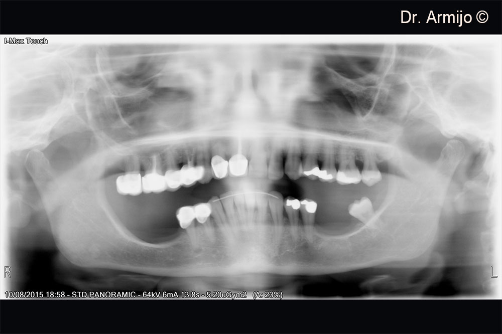
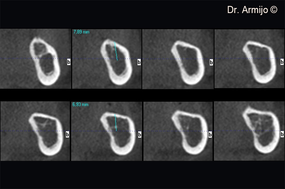
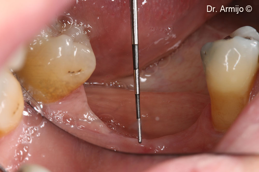
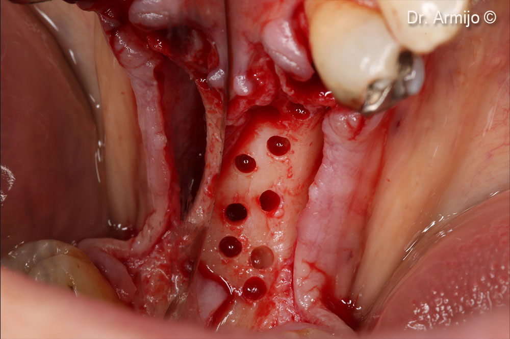
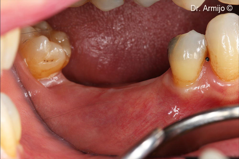
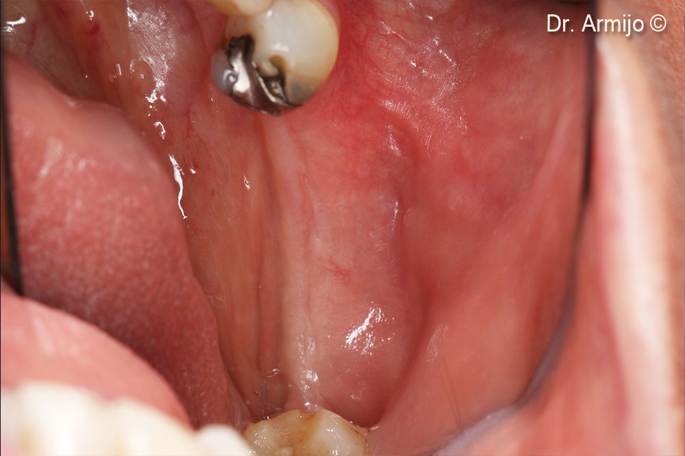
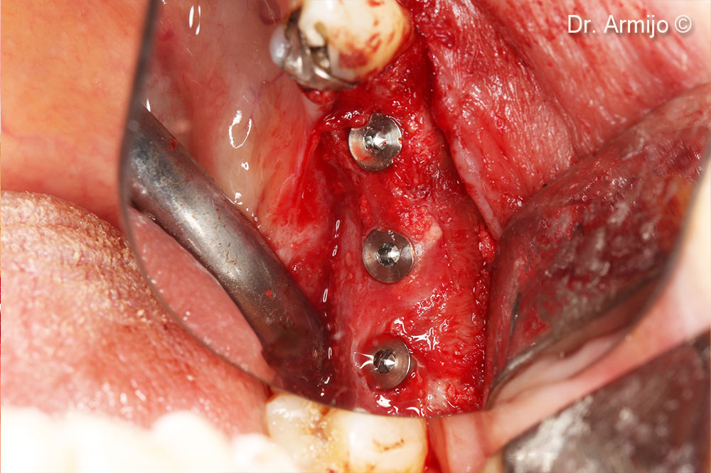
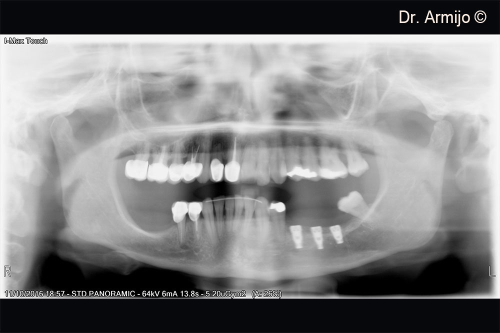
Initial situation
A female patient (60 years old) shows a horizontal/vertical defect in the lower jaw
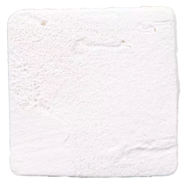
OsteoBiol by Tecnoss
Attention please! The OsteoBiol® website contains information on Medical Devices, which may be dangerous for the patient health and safety if not used exclusively by medical professionals.