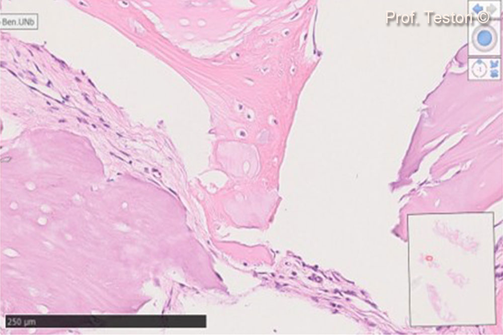
Treatment of a complex defect in the upper jaw: the F.I.R.S.T. protocol
Prof Tiziano Testori
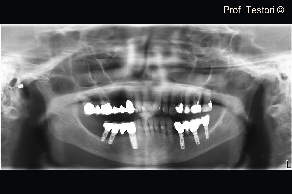
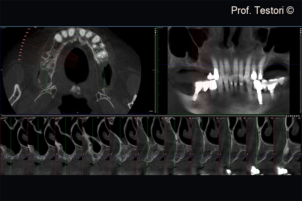
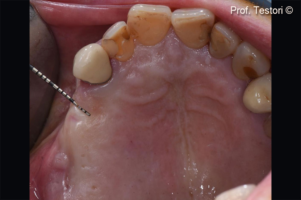
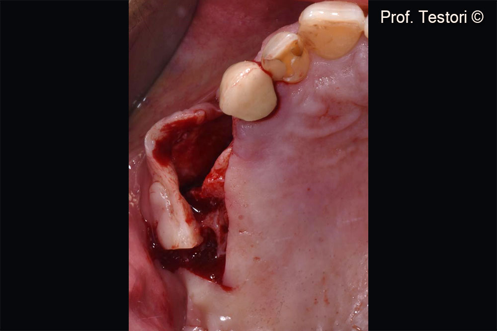
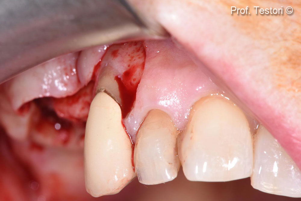
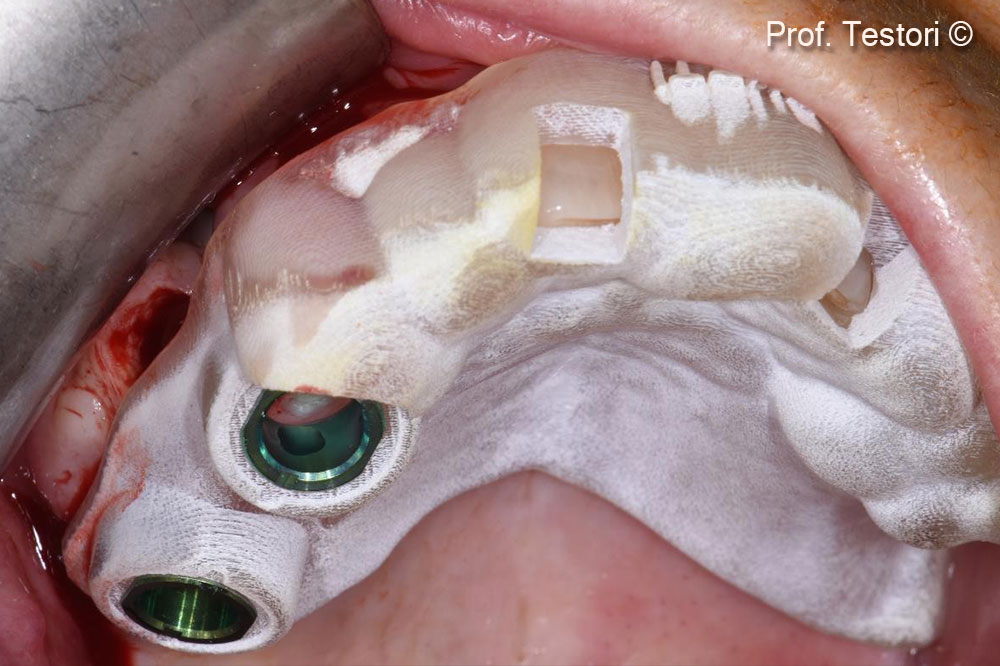
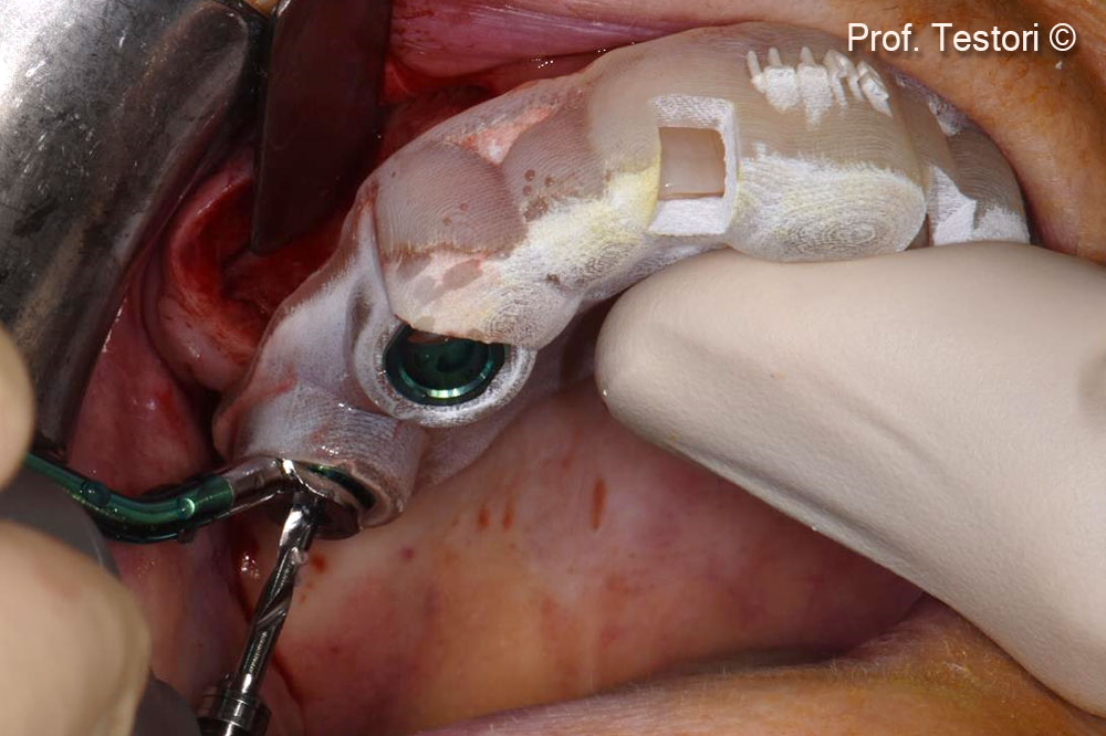
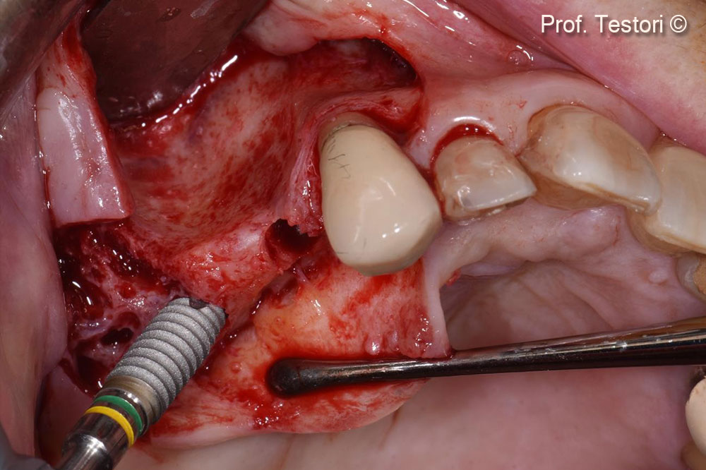
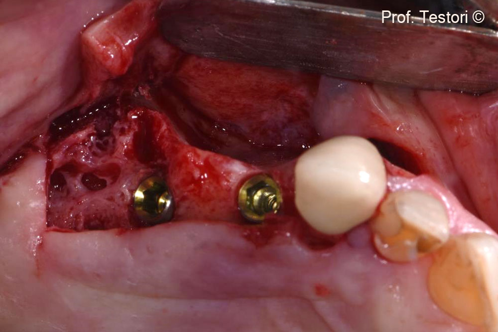
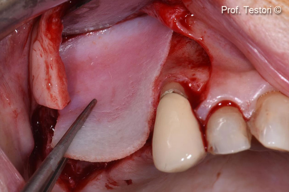
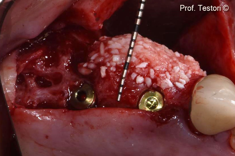
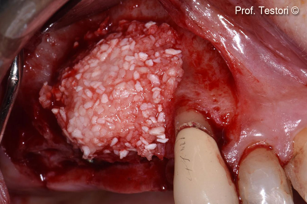
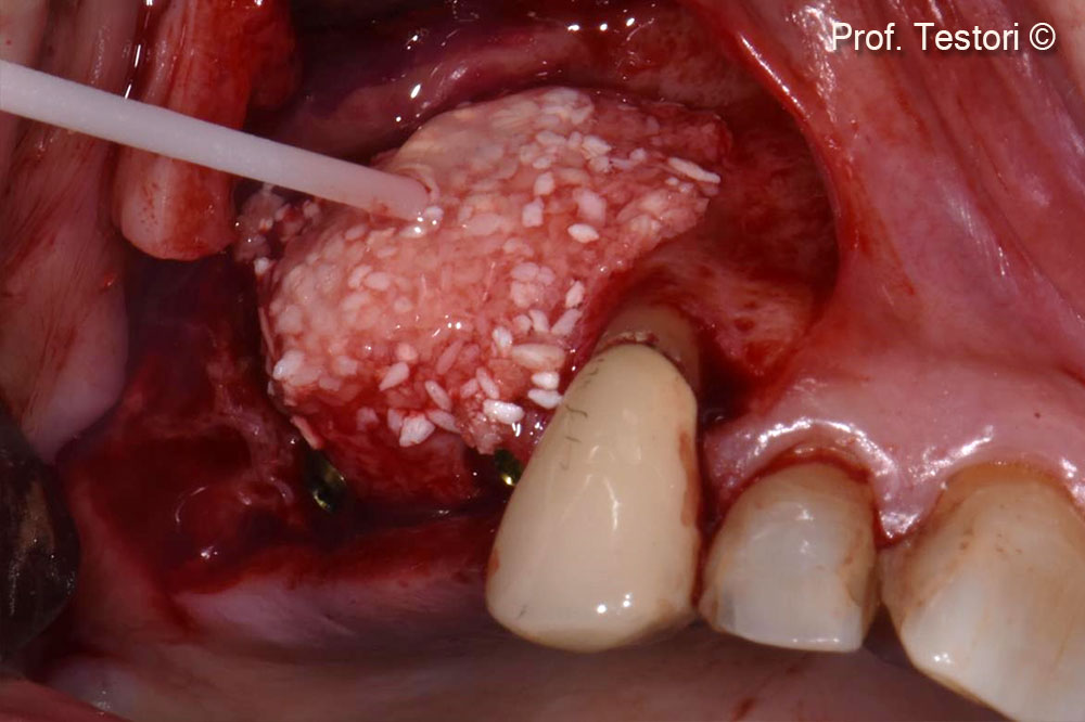
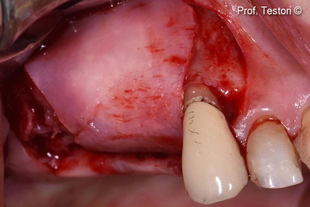
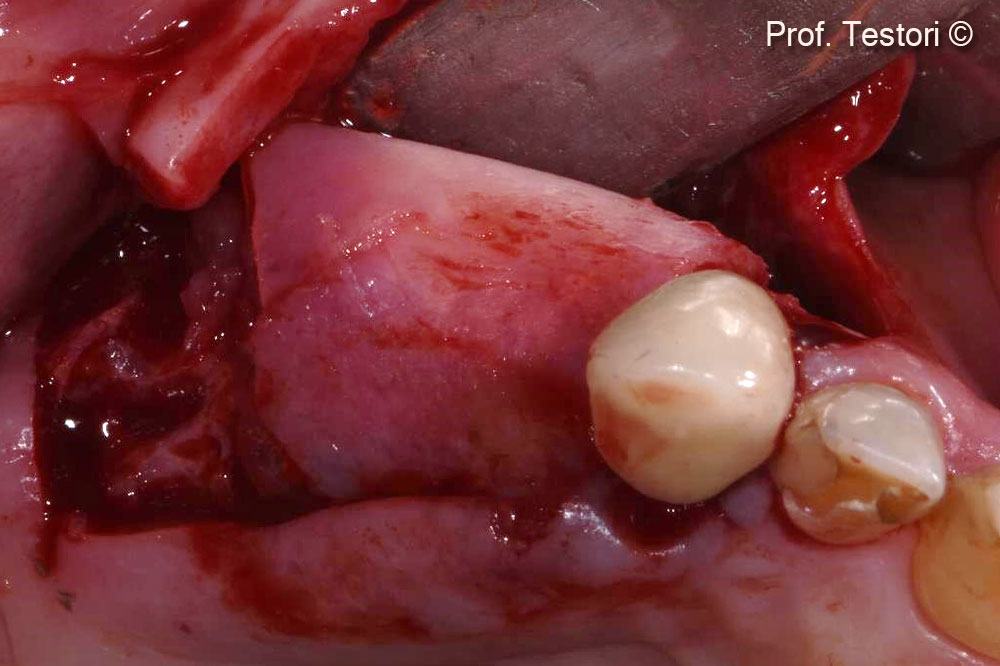
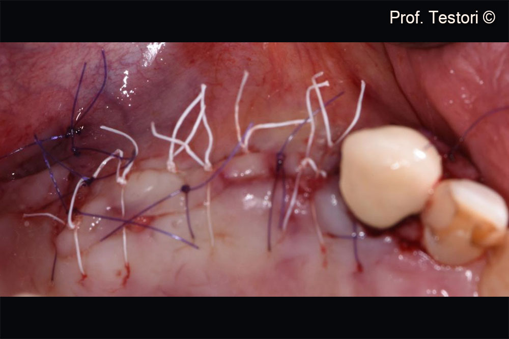
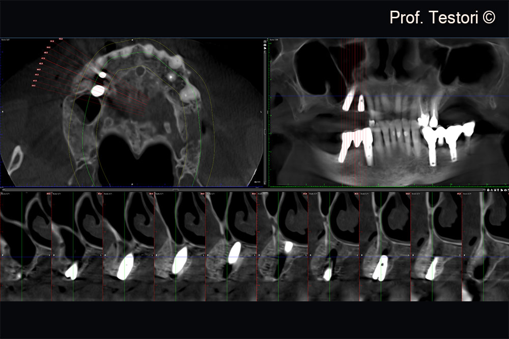
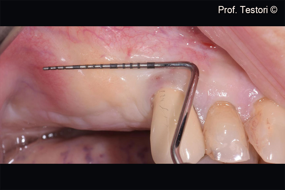
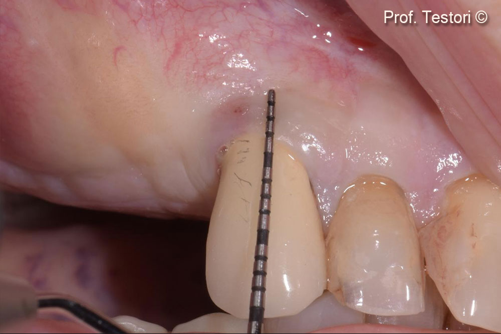
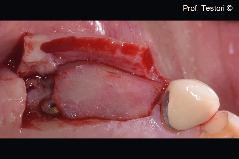
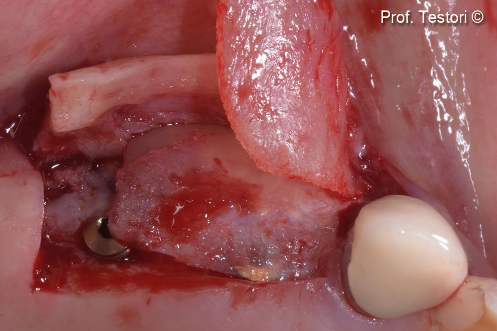
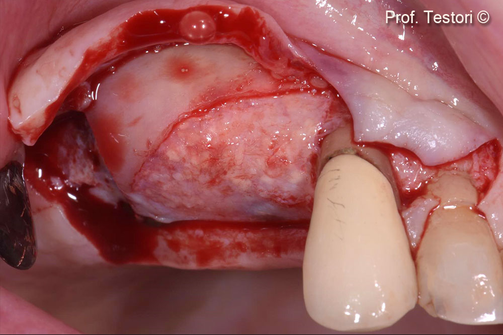
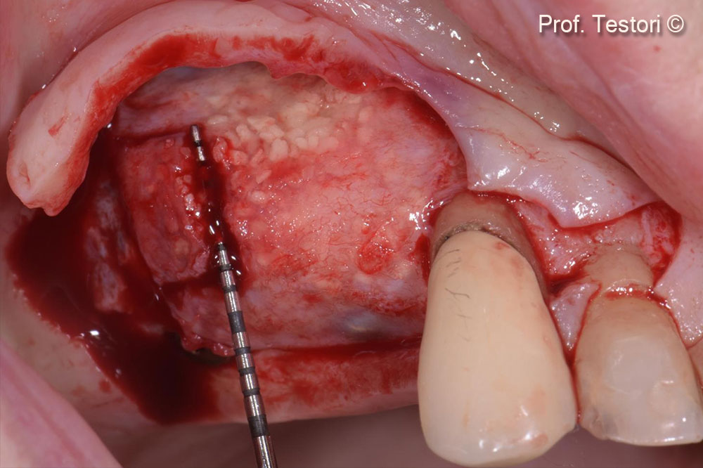
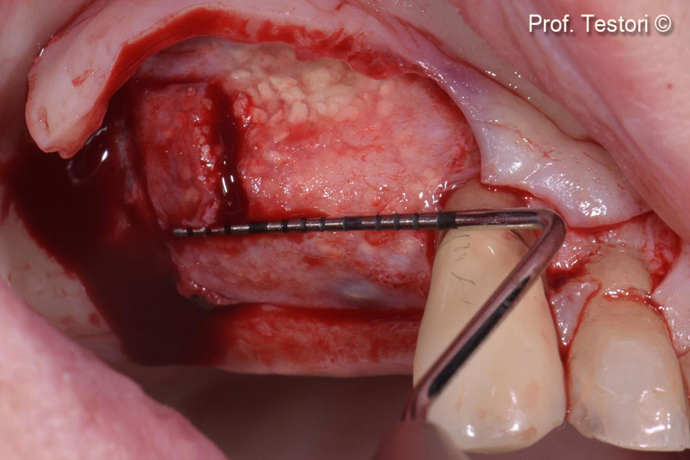
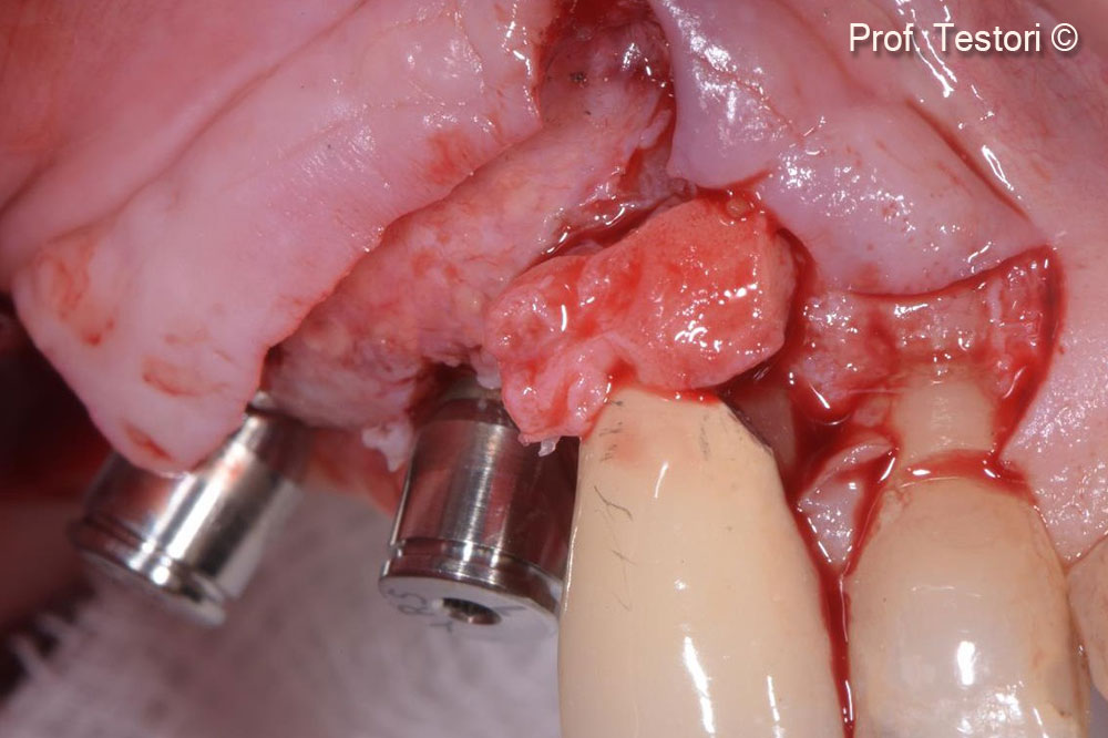
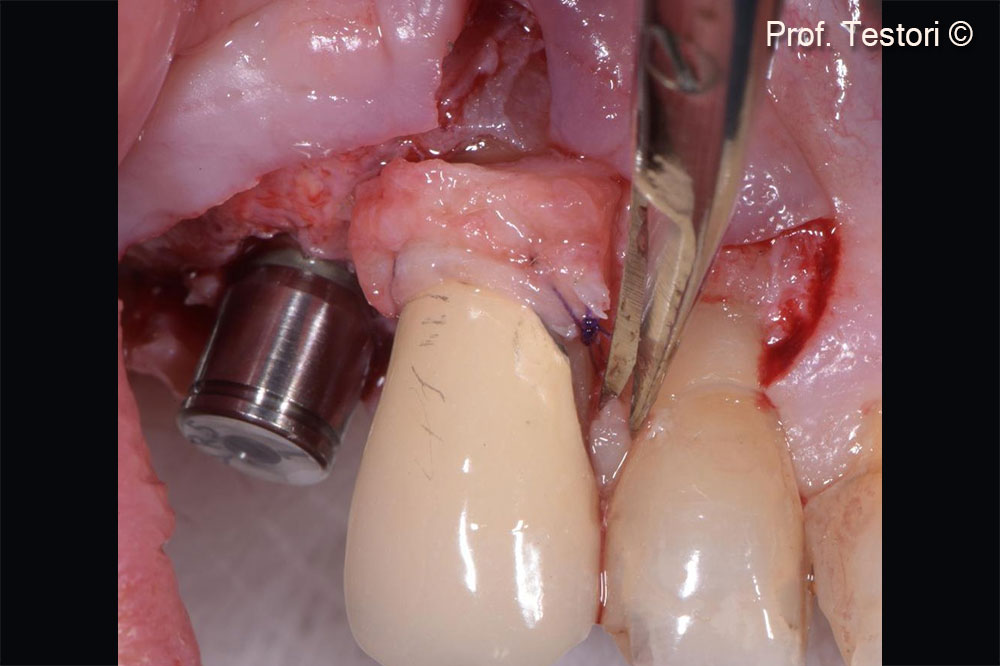
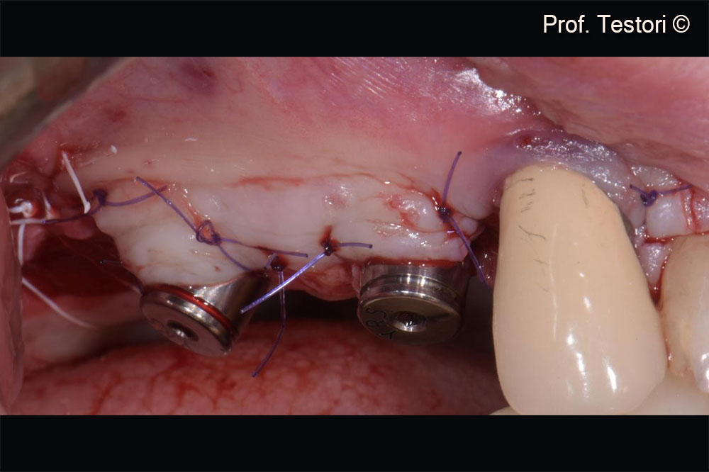
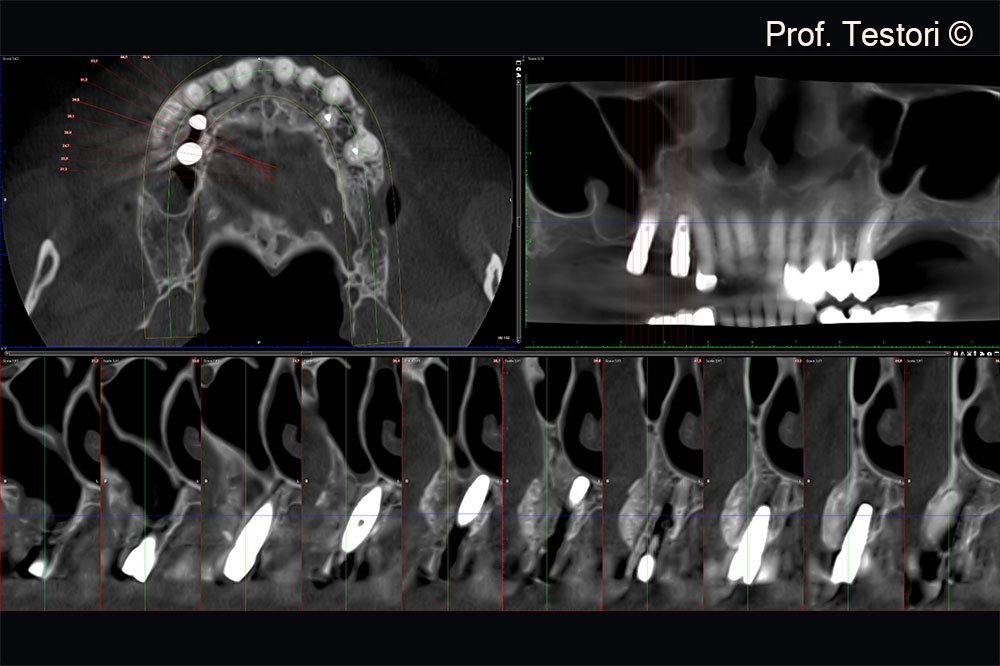
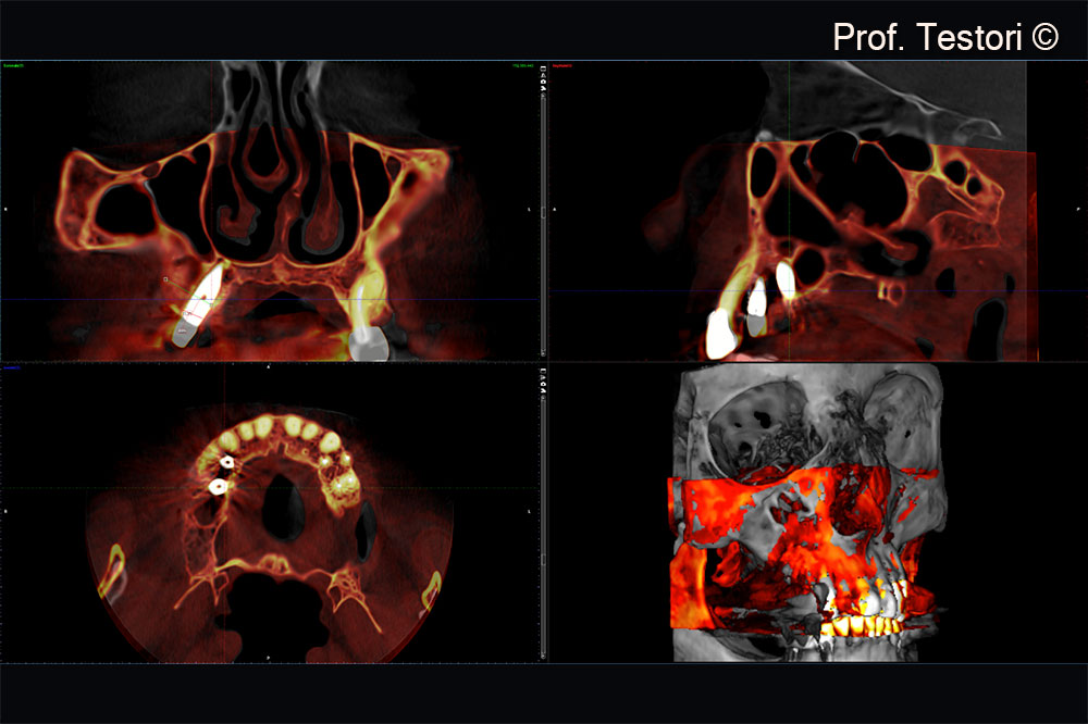
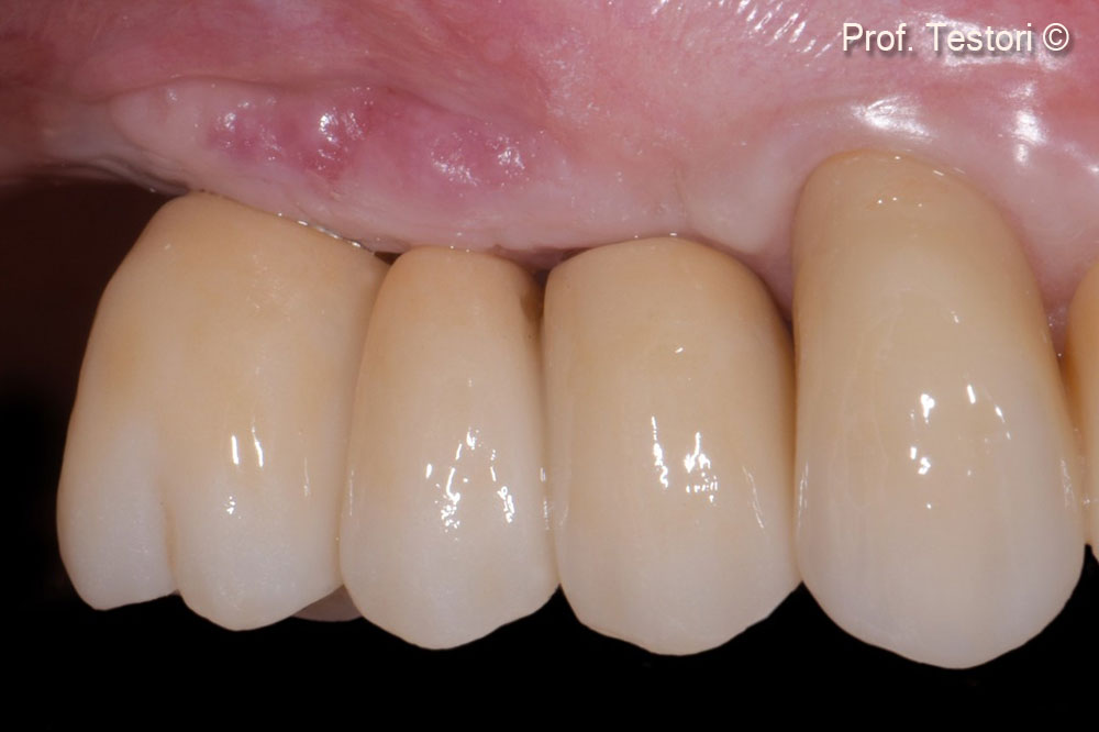
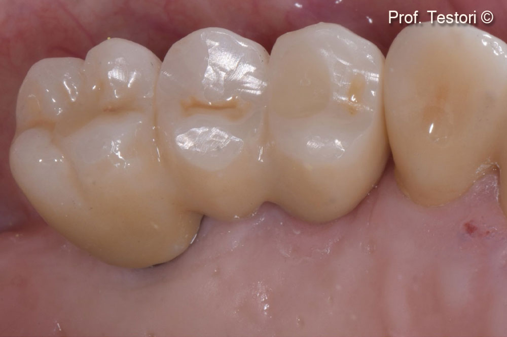
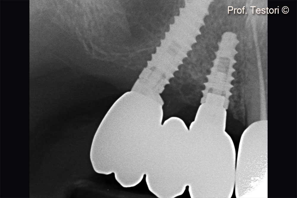
Initial situation
A female patient (62 years old) requires rehabilitation of the upper jaw.
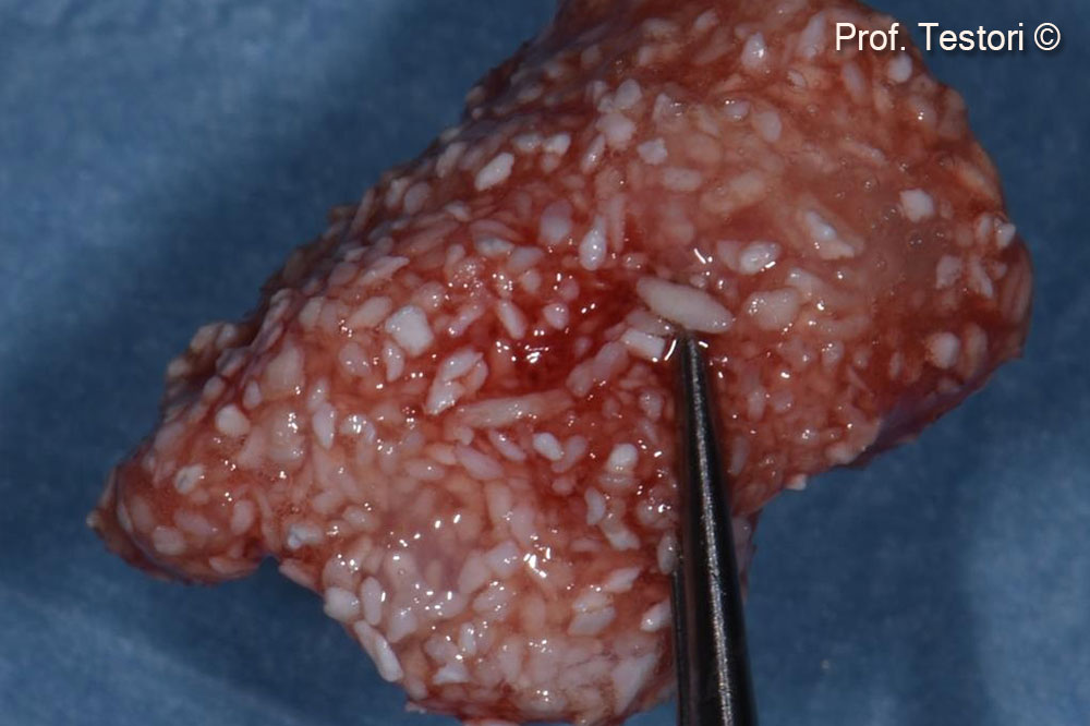
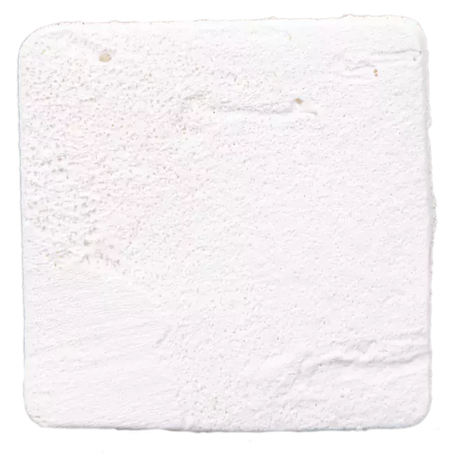
OsteoBiol by Tecnoss
Attention please! The OsteoBiol® website contains information on Medical Devices, which may be dangerous for the patient health and safety if not used exclusively by medical professionals.