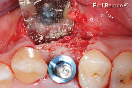
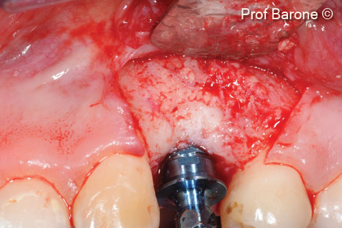
Ridge augmentation in the upper jaw

Prof. Antonio Barone
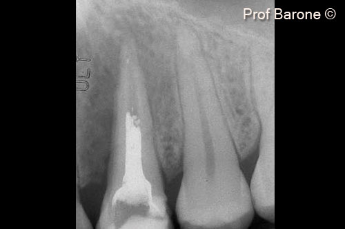
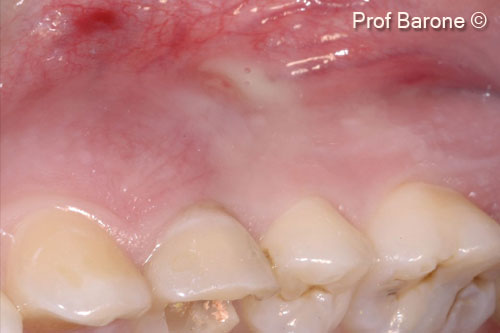
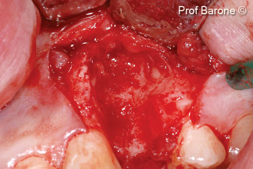
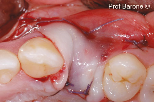
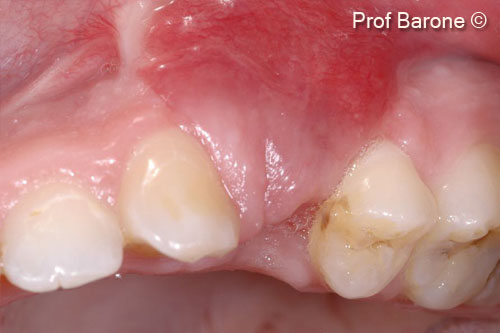
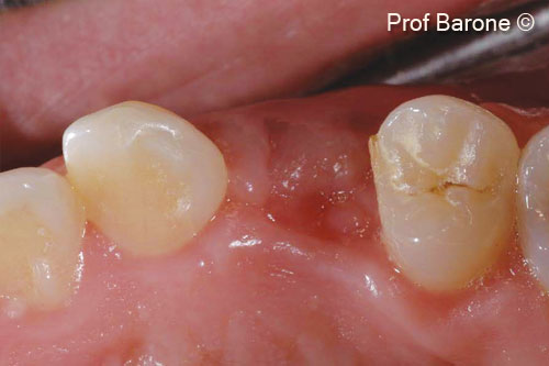
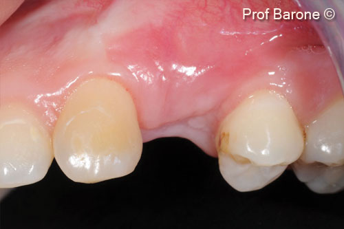
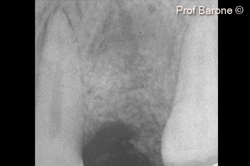
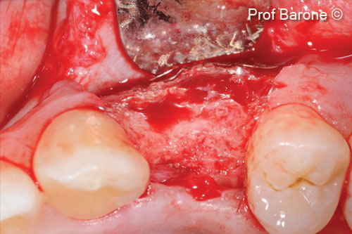
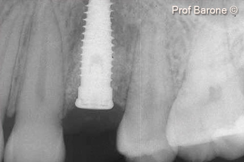
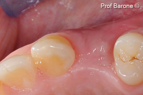
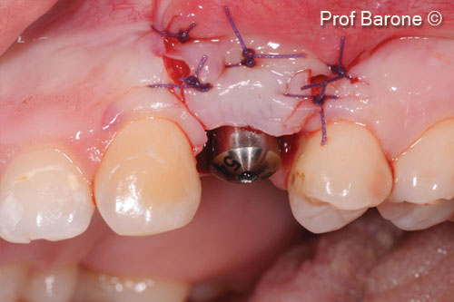
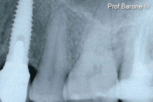
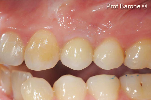
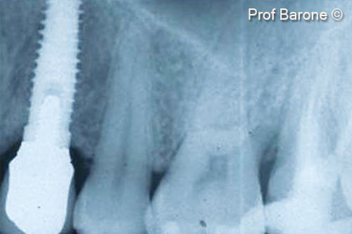
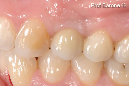
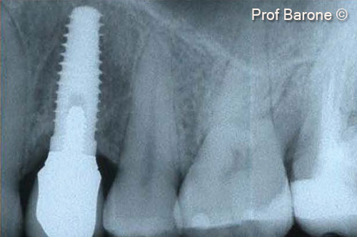
Initial situation

A female patient (27 years old) shows acute infection and suppuration from a buccal fistula
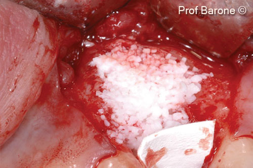
Used biomaterials
  • mp3®
  • Evolution
1. Pre-operative periapical radiograph tooth #24

1. Pre-operative periapical radiograph tooth #24

2. Clinical view. Acute infection, suppuration from a buccal fistula, mesial and distal probing depth 12mm, buccal probing depth 15 mm and mobility degree 3

2. Clinical view. Acute infection, suppuration from a buccal fistula, mesial and distal probing depth 12mm, buccal probing depth 15 mm and mobility degree 3

3. Intra-operative view, full thickness flap was elevated, the tooth was extracted and a severe bone defect was present

3. Intra-operative view, full thickness flap was elevated, the tooth was extracted and a severe bone defect was present

4. The bone defect was grafted with OsteoBiol® mp3® and the graft protected with an OsteoBiol® Evolution membrane

4. The bone defect was grafted with OsteoBiol® mp3® and the graft protected with an OsteoBiol® Evolution membrane

5. Primary soft tissue closure was obtained

5. Primary soft tissue closure was obtained

6. Soft tissue healing 6 weeks after the ridge augmentation procedure. Buccal view

6. Soft tissue healing 6 weeks after the ridge augmentation procedure. Buccal view

7. Soft tissue healing 6 weeks after the ridge augmentation procedure. Occlusal view

7. Soft tissue healing 6 weeks after the ridge augmentation procedure. Occlusal view

8. Soft tissue healing 6 months after the ridge augmentation procedure, the muco-gingival junction was displaced coronally. Buccal view

8. Soft tissue healing 6 months after the ridge augmentation procedure, the muco-gingival junction was displaced coronally. Buccal view

9. Periapical x-ray of tooth #24 6 months after ridge augmentation procedure

9. Periapical x-ray of tooth #24 6 months after ridge augmentation procedure

10. Bone regeneration 6 months after the ridge augmentation procedure, some residual particles are still visible, the biomaterial granules were well integrated in the new and well vascularized bone

10. Bone regeneration 6 months after the ridge augmentation procedure, some residual particles are still visible, the biomaterial granules were well integrated in the new and well vascularized bone

11. Implant placement in a prosthetically driven position, occlusal view

11. Implant placement in a prosthetically driven position, occlusal view

12. Implant placement, buccal view. The newly formed bone appeared well vascularized

12. Implant placement, buccal view. The newly formed bone appeared well vascularized

13. Peri-apical radiograph after implant placement

13. Peri-apical radiograph after implant placement

14. Soft tissue healing 3 months after implant placement, tooth position #24

14. Soft tissue healing 3 months after implant placement, tooth position #24

15. Second stage of surgery, apically positioned flap to rebuild a correct soft tissue architecture

15. Second stage of surgery, apically positioned flap to rebuild a correct soft tissue architecture

16. Final restoration

16. Final restoration

17. Final restoration

17. Final restoration

18. 7 years follow-up

18. 7 years follow-up

19. 7 years follow-up

19. 7 years follow-up

20. 13 years follow-up

20. 13 years follow-up

21. 13 years follow-up

21. 13 years follow-up

Used biomaterials
mp3
mp3®
evolution
Evolution
Are you a medical professional?
No

Attention please! The OsteoBiol® website contains information on Medical Devices, which may be dangerous for the patient health and safety if not used exclusively by medical professionals.