Clinical case of bilateral sinus lift for implant placement
Dr. Raquel Zita Gomes
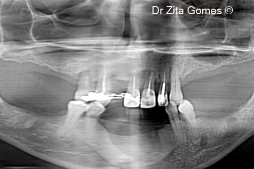
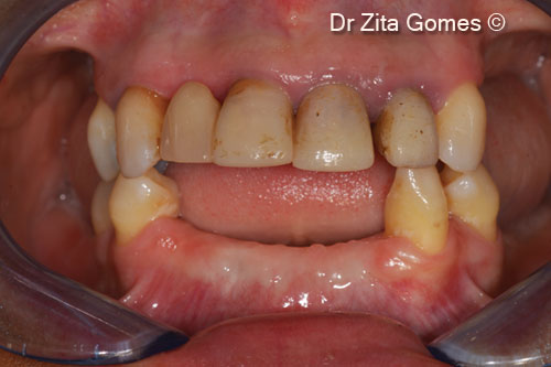
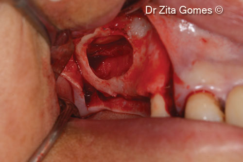
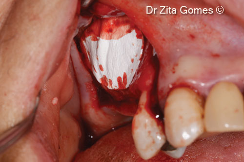
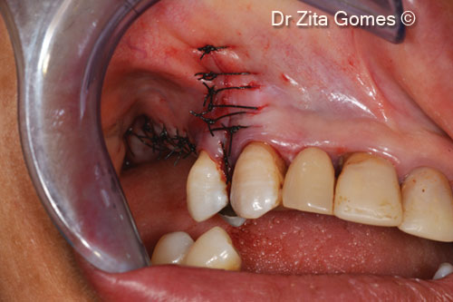
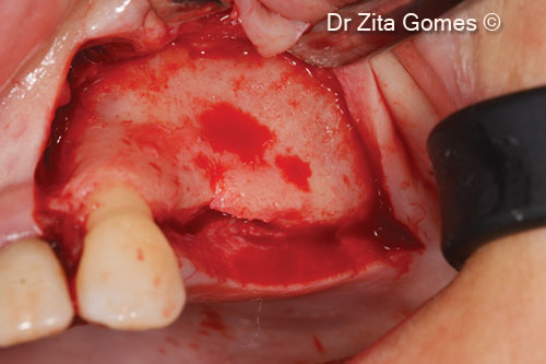
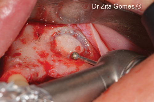
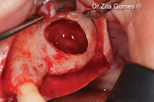
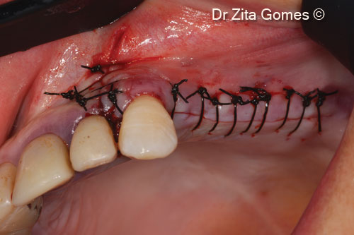
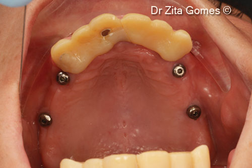
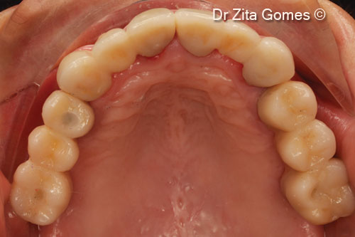
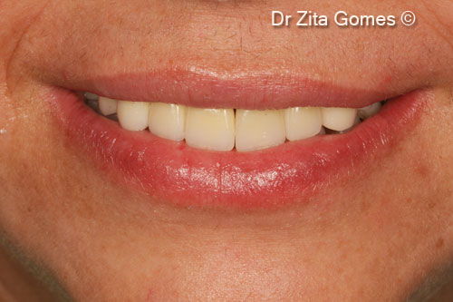
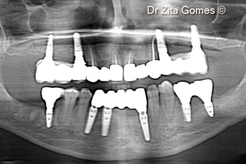
Initial situation
A female patient (50 years old) shows pneumatization of both maxillary sinuses
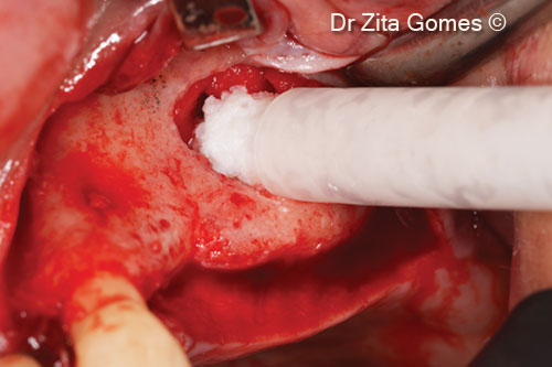
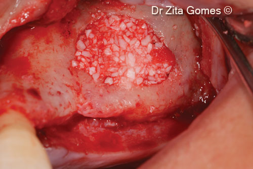
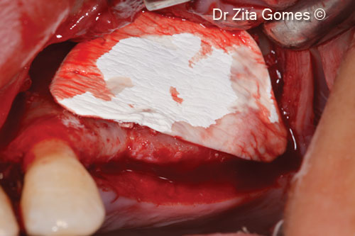
OsteoBiol by Tecnoss
Attention please! The OsteoBiol® website contains information on Medical Devices, which may be dangerous for the patient health and safety if not used exclusively by medical professionals.