Vertical Guided Bone and Tissue Regeneration with F.I.R.S.T.
Dr Vincenzo Foti
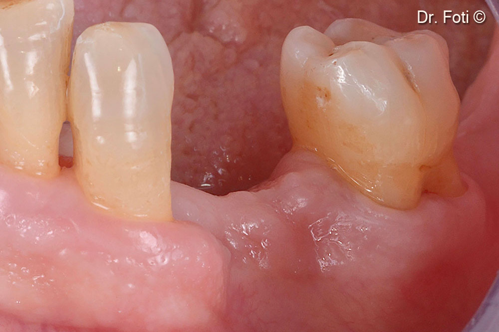
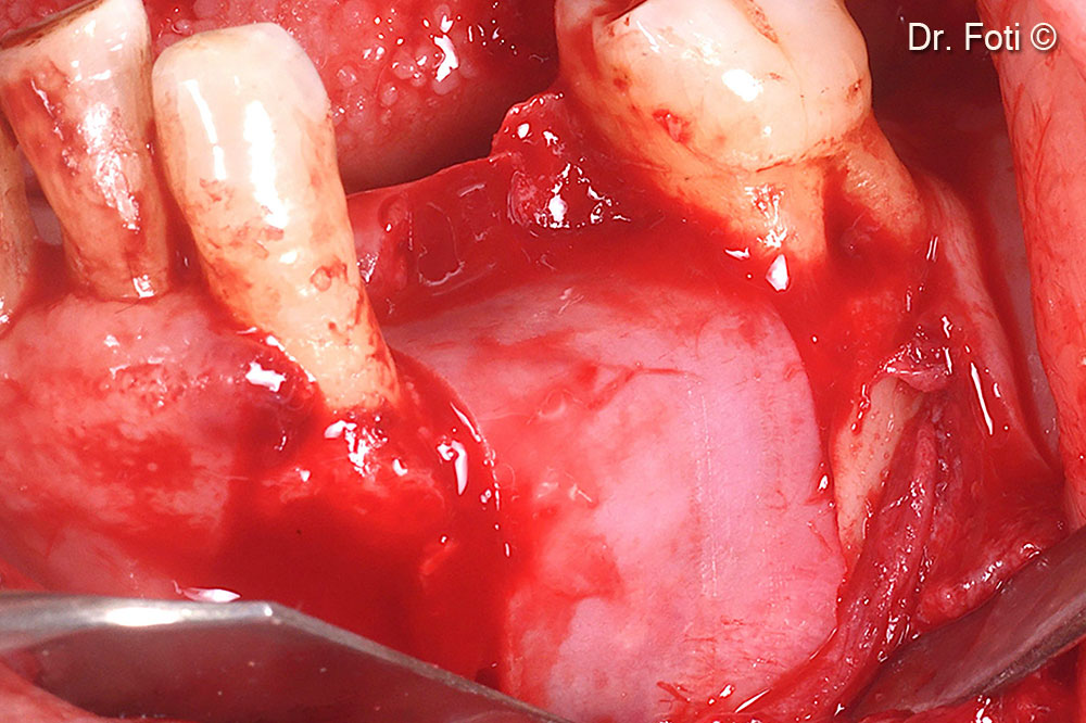
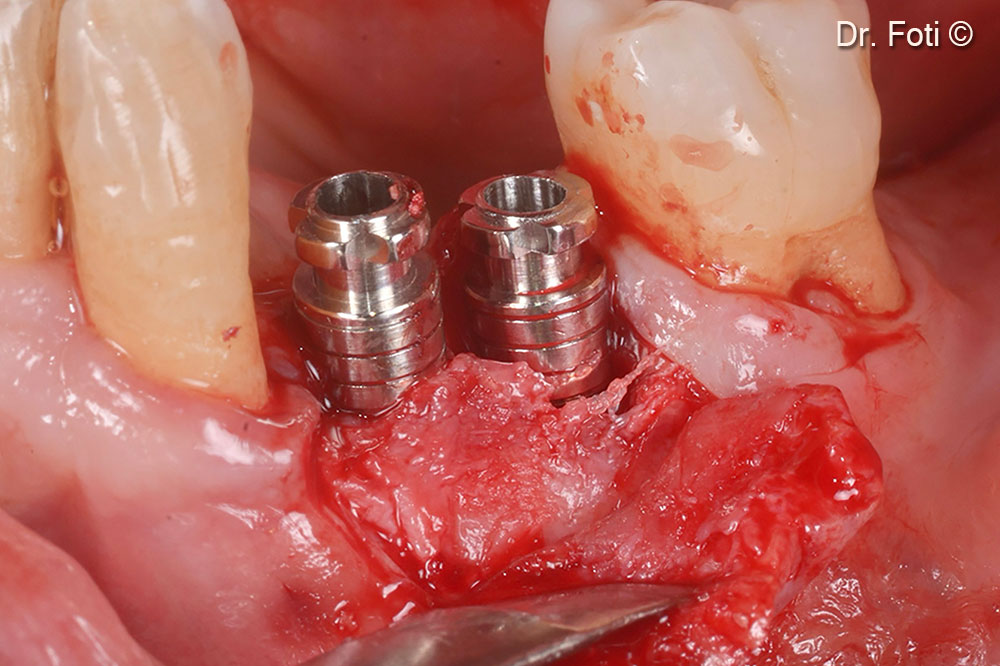
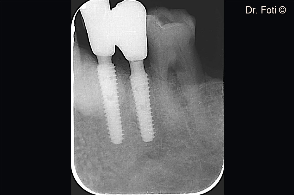
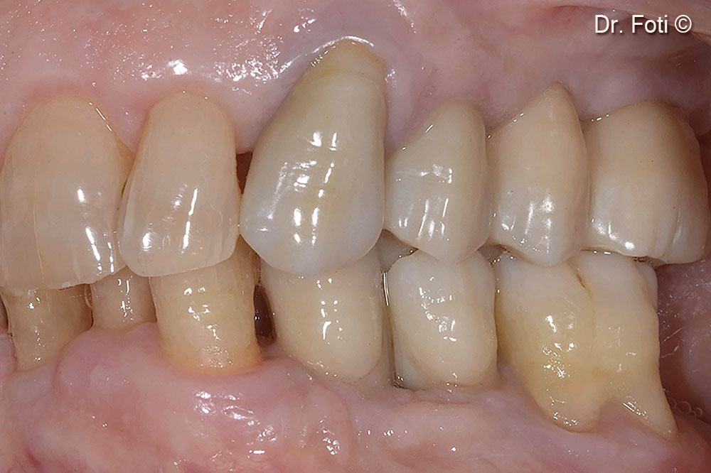
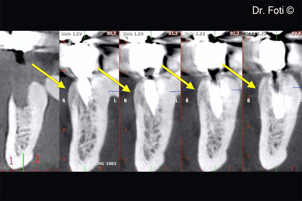
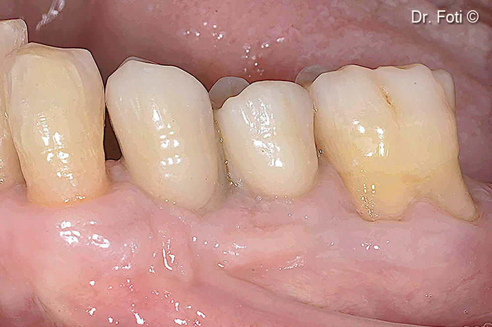
Initial situation
A male patient (71 years old) lacks teeth in positions 34 and 35.
OsteoBiol by Tecnoss
Attention please! The OsteoBiol® website contains information on Medical Devices, which may be dangerous for the patient health and safety if not used exclusively by medical professionals.