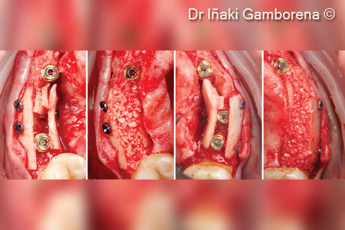
Vertical and horizontal reconstruction of the esthetic area
Dr. Iñaki Gamborena
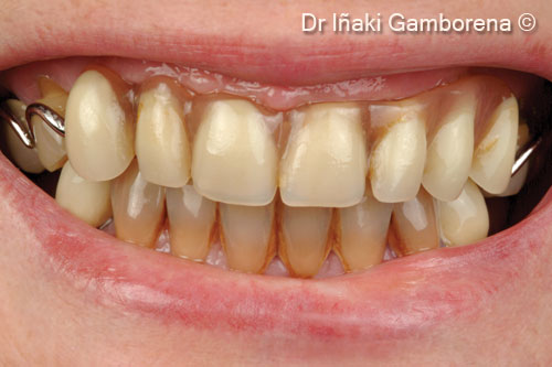
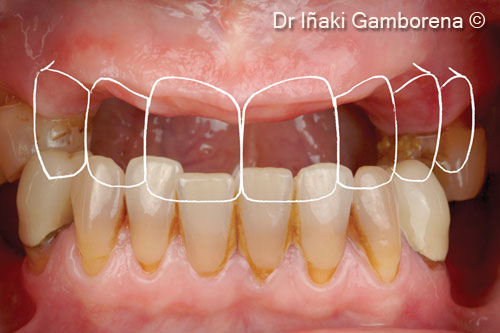
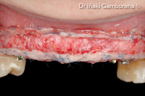
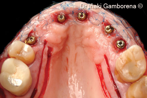
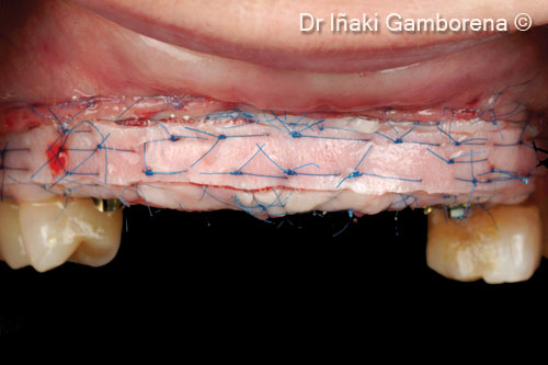
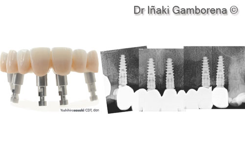
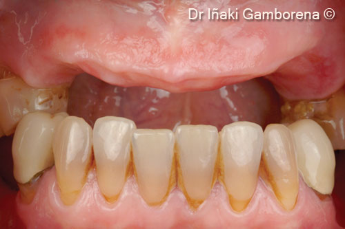
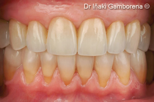
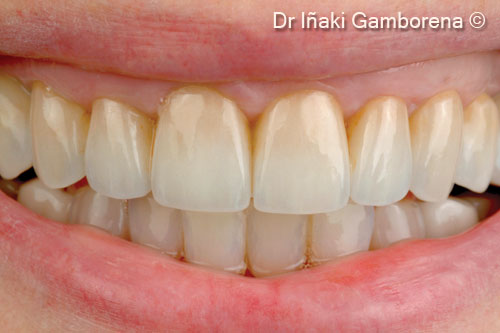
Initial situation
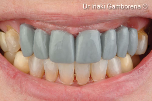
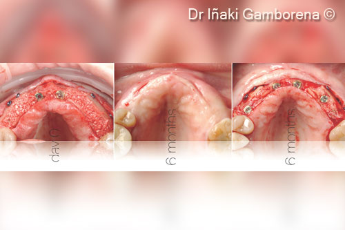
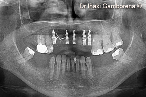
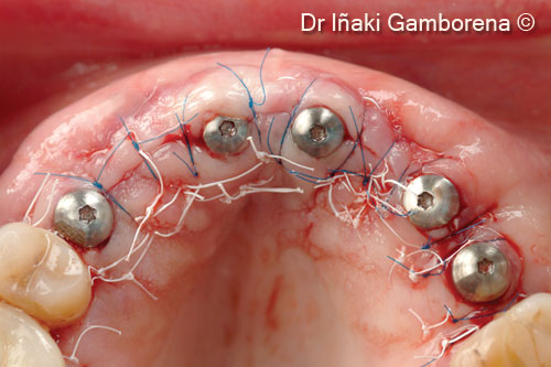
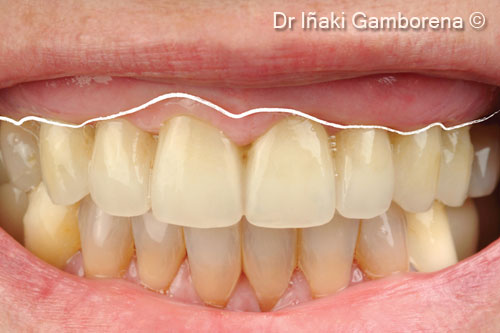
A female patient (53 years old) wishes to have a fixed upper restoration on implants
OsteoBiol by Tecnoss
Attention please! The OsteoBiol® website contains information on Medical Devices, which may be dangerous for the patient health and safety if not used exclusively by medical professionals.