Initial situation
A patient reports a gingival recession and a fistula.
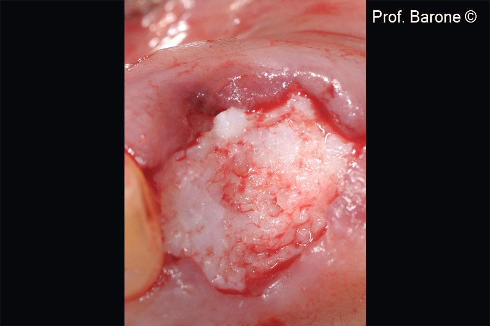
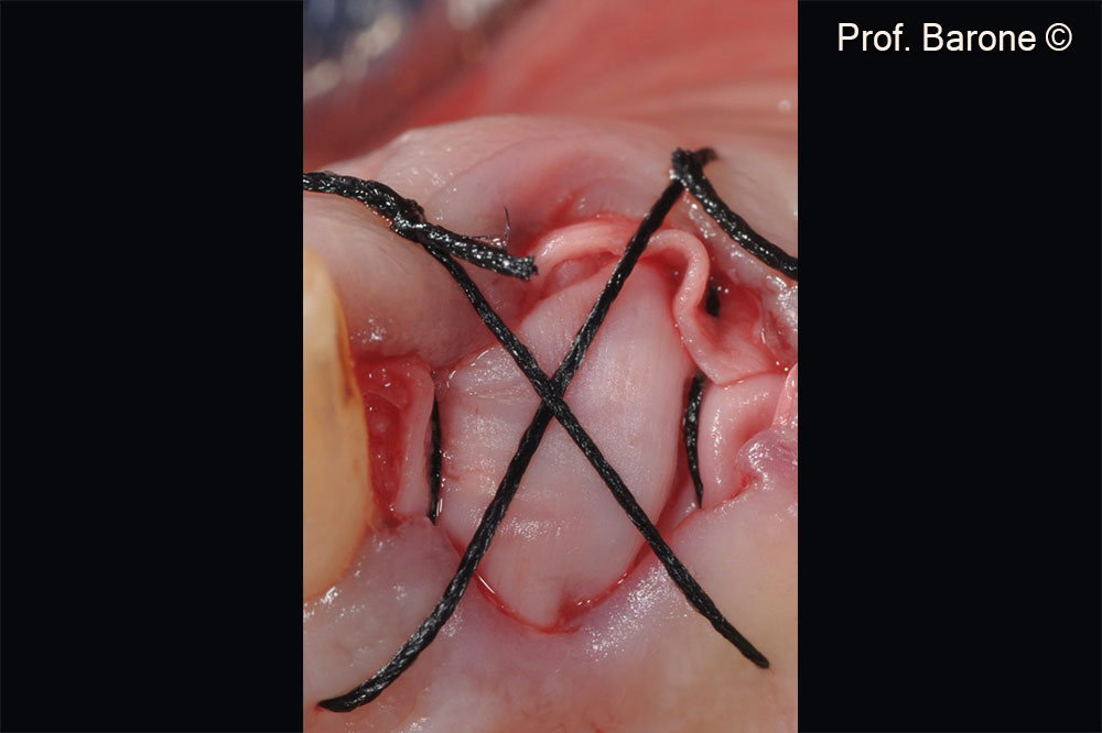
Used biomaterials
- GTO®
- Evolution
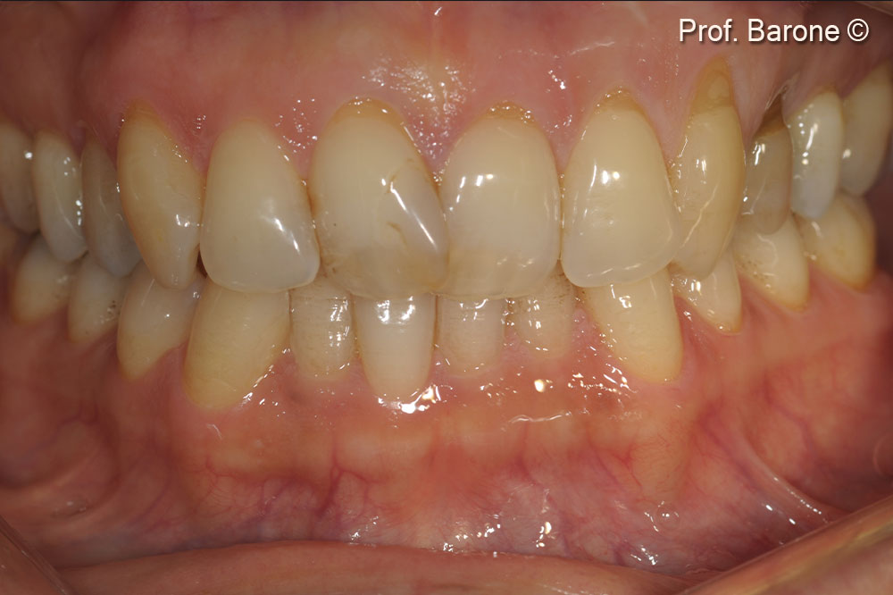
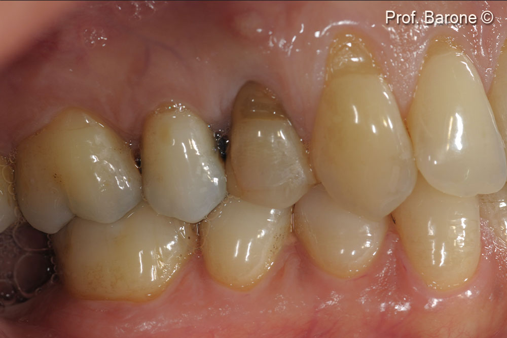
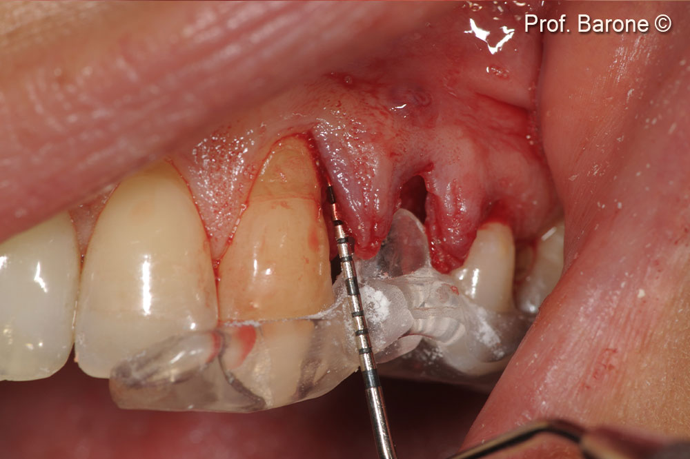
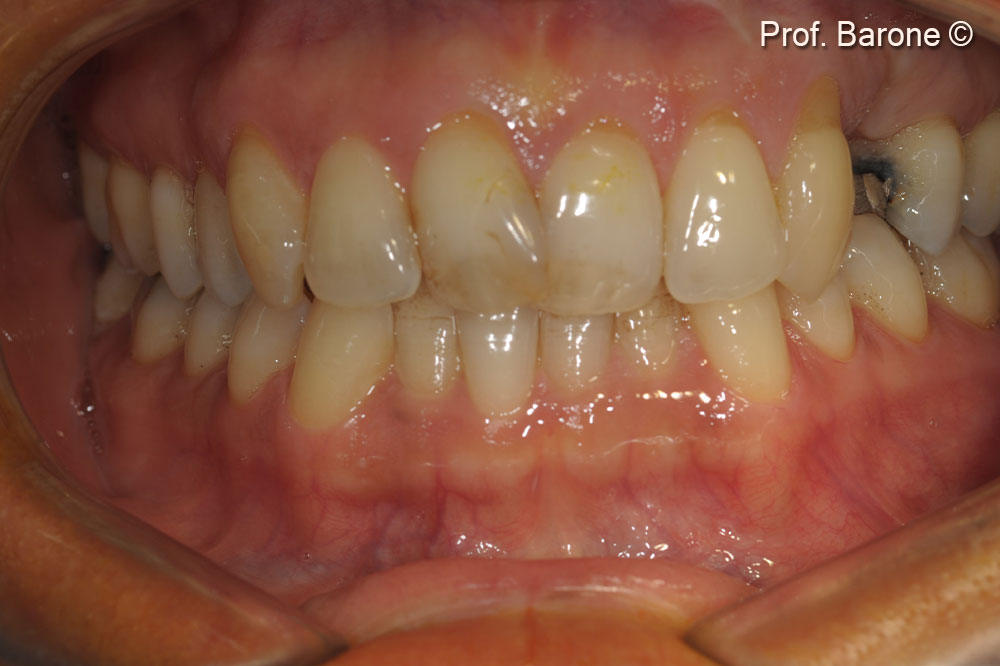
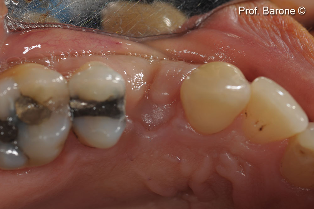

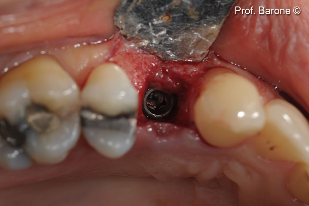
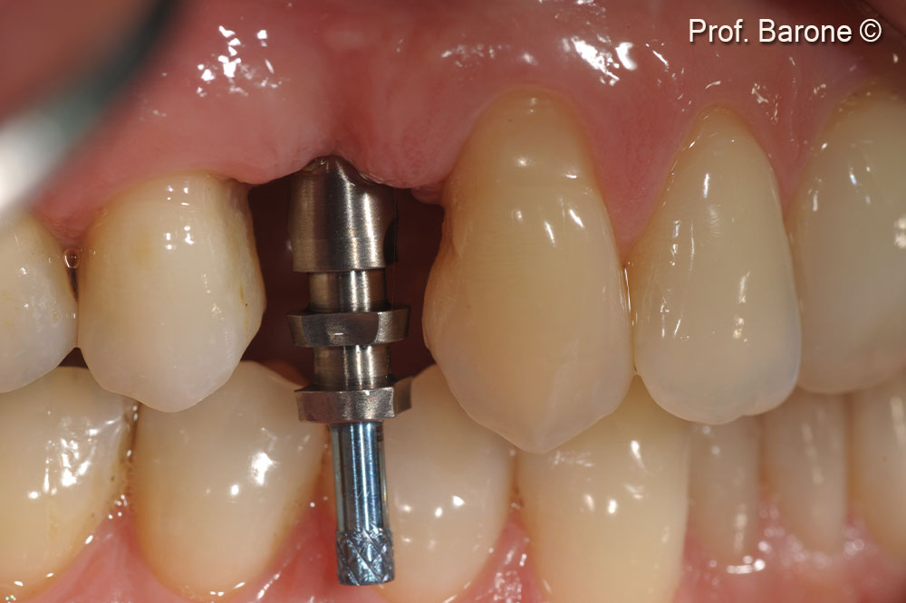
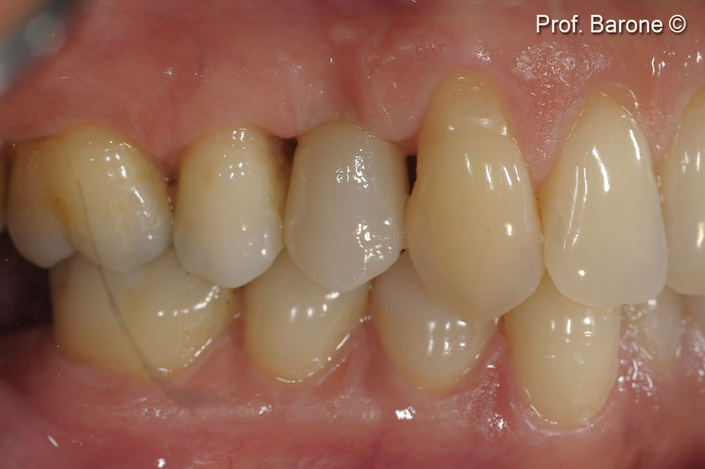
A patient reports a gingival recession and a fistula.
Attention please! The OsteoBiol® website contains information on Medical Devices, which may be dangerous for the patient health and safety if not used exclusively by medical professionals.