Sinus augmentation with simultaneous horizontal GBR
Dr. Sorin Andreica
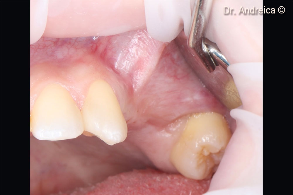
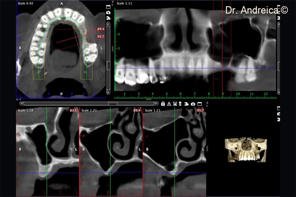
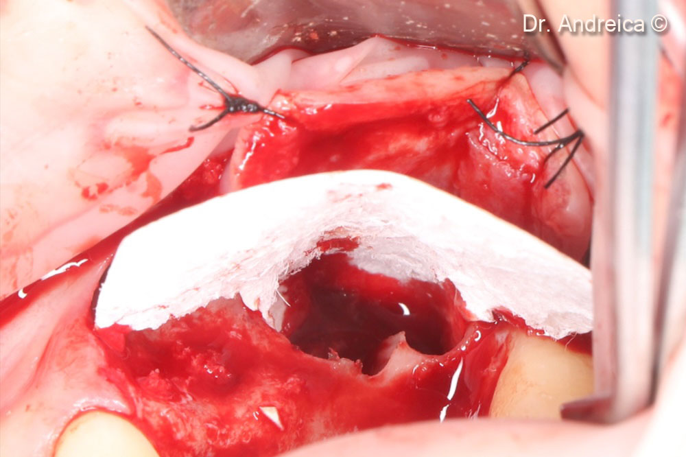
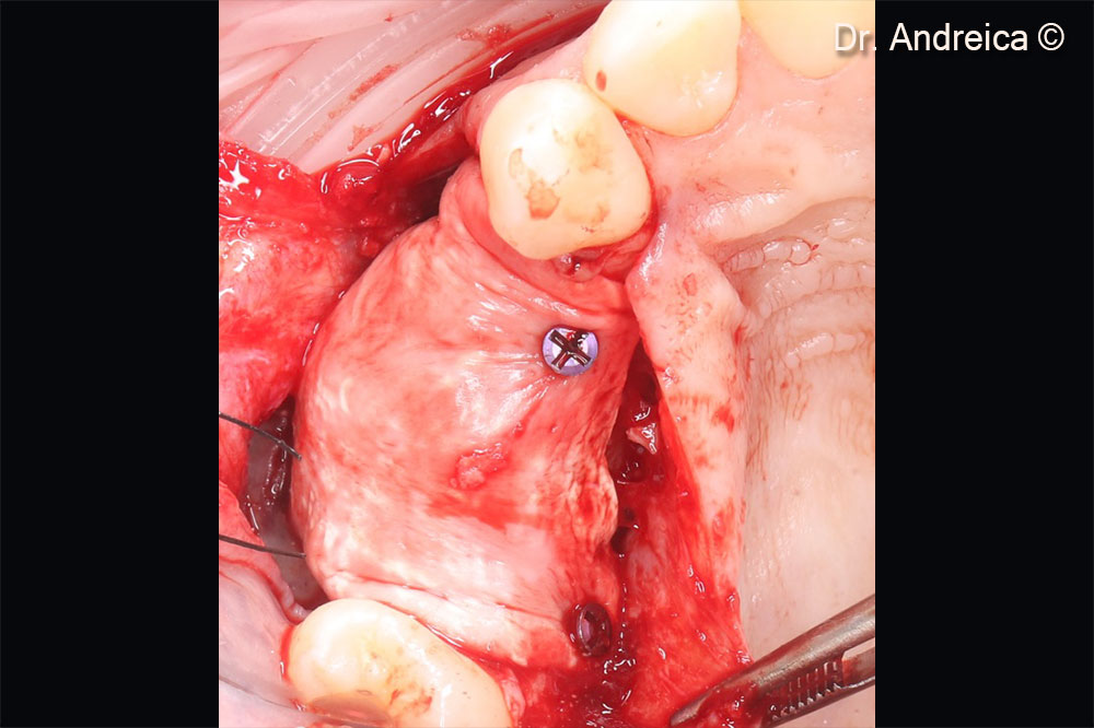
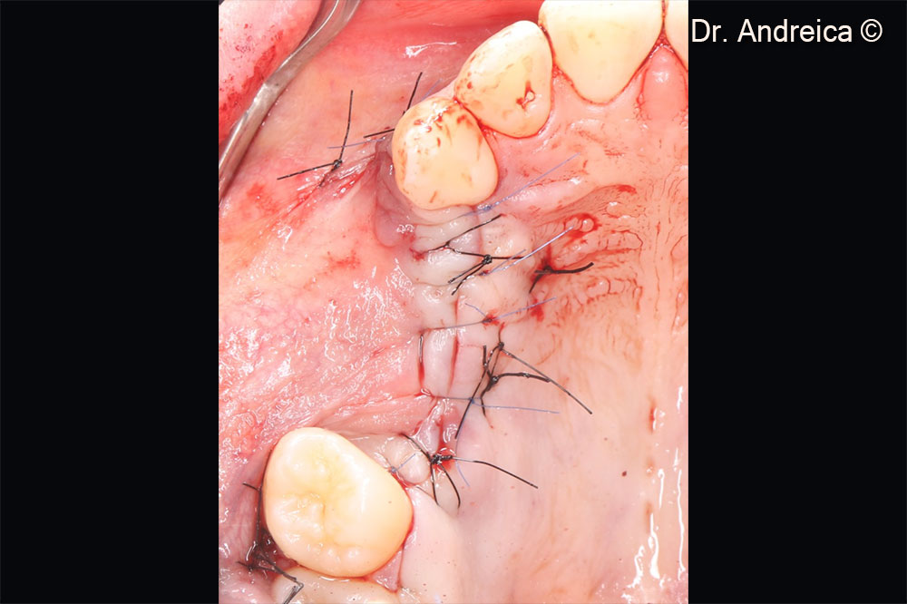
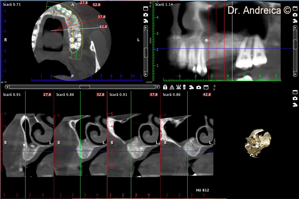
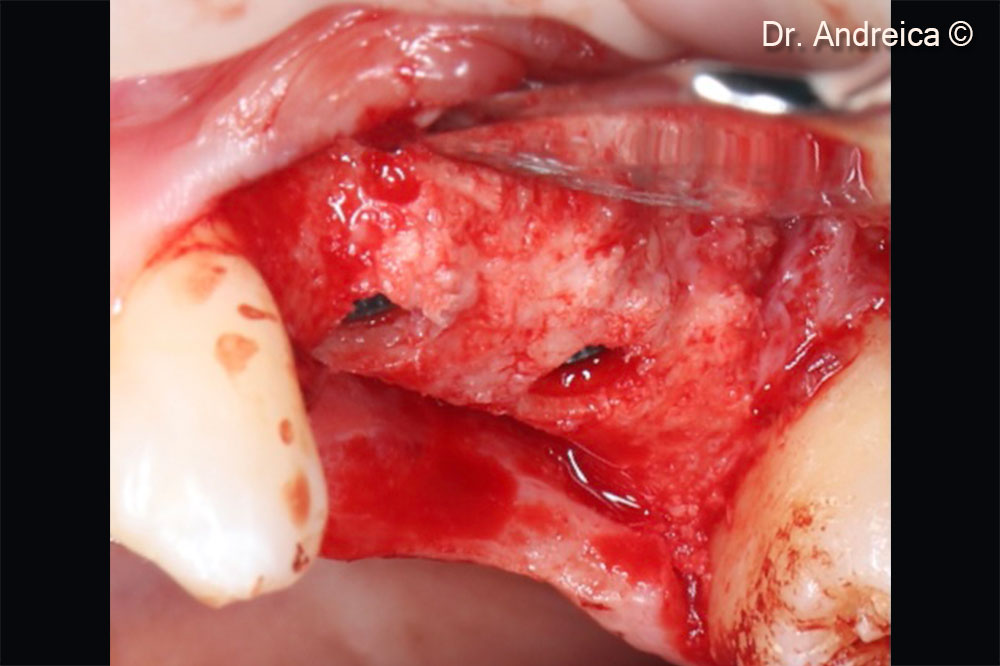
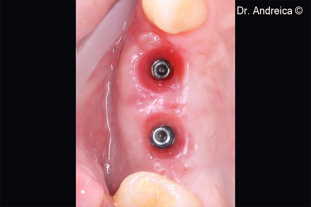
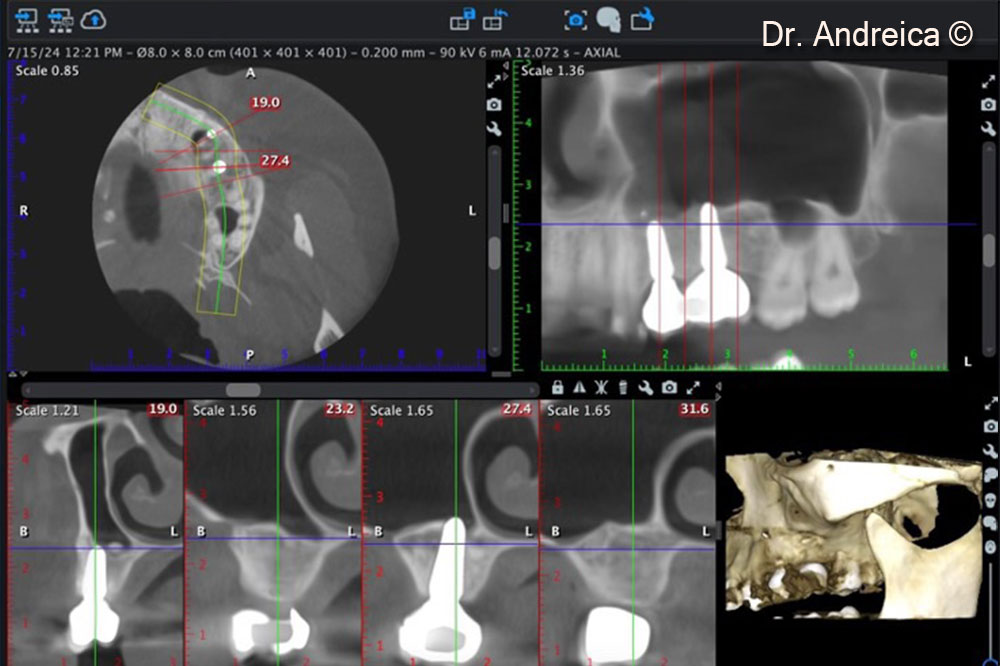
Initial situation
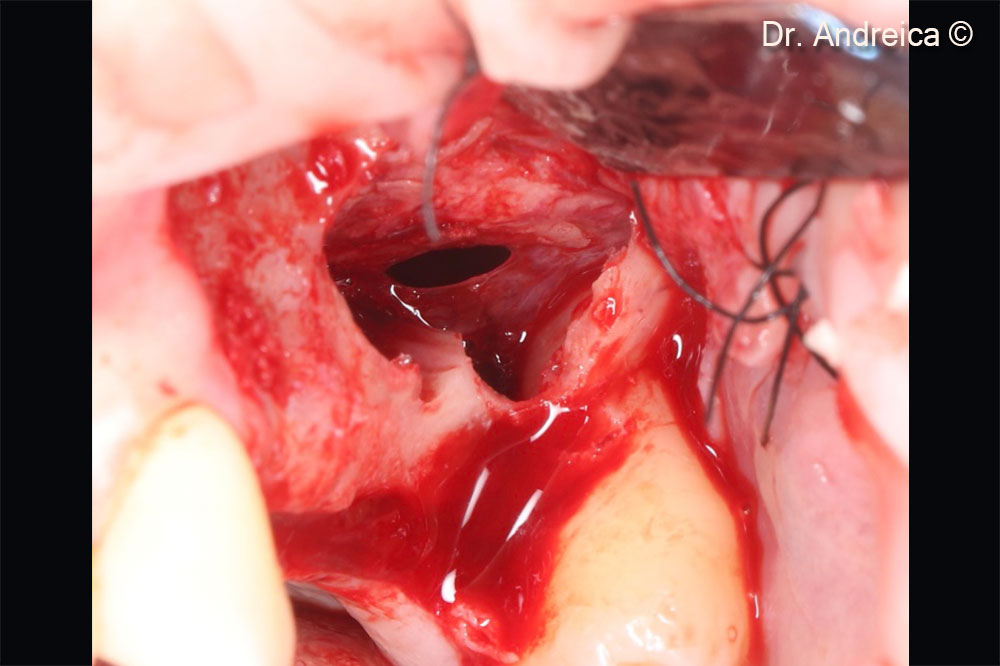
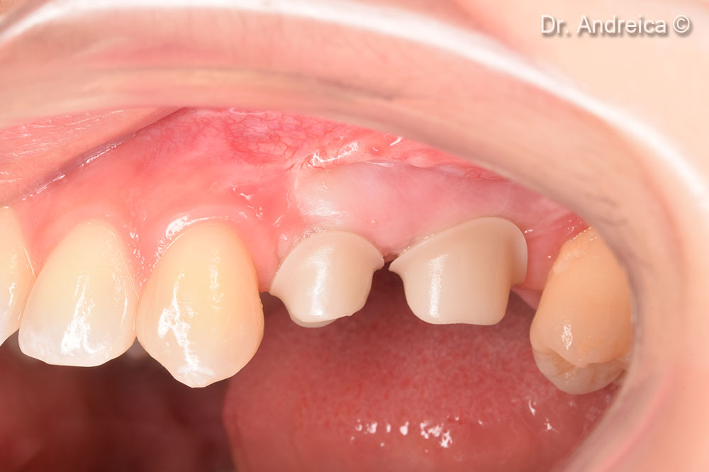
A female patient (35 years old) shows a horizontal defect in the posterior area of the jaw.
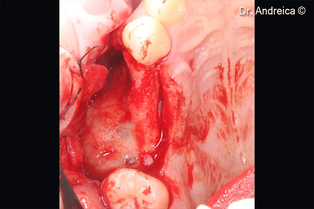
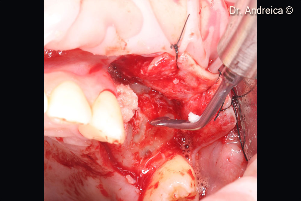
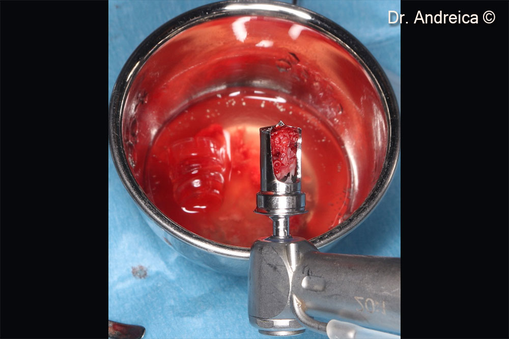
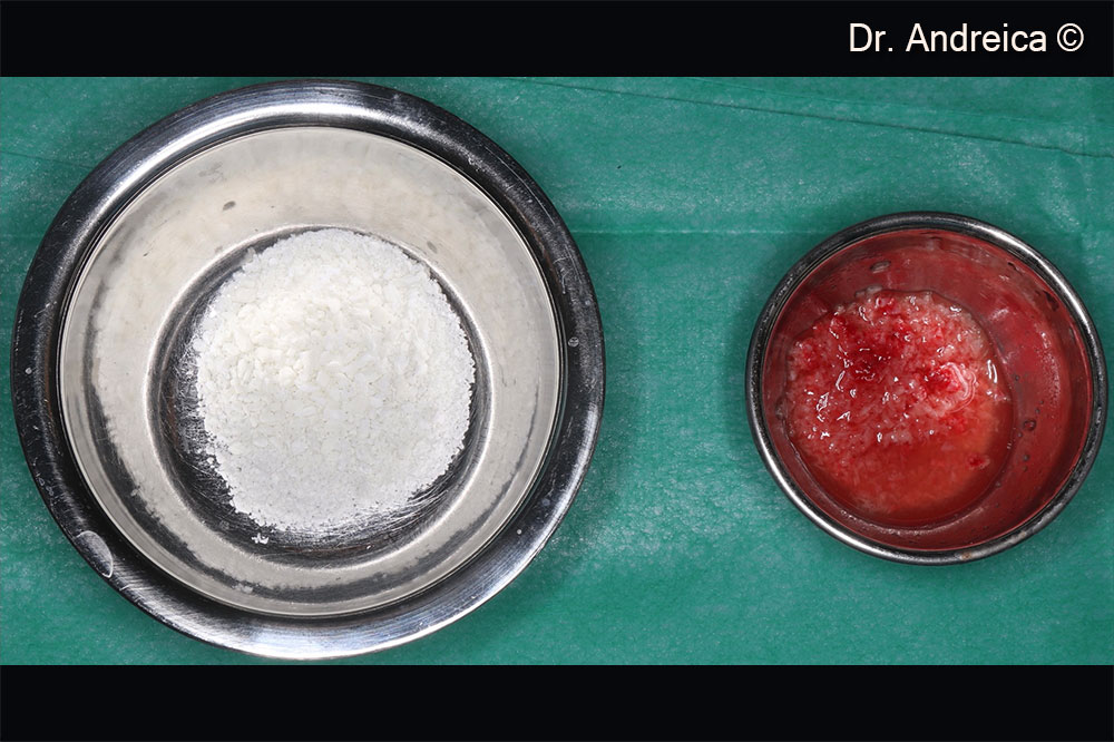
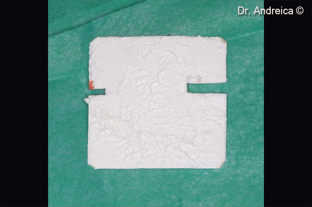
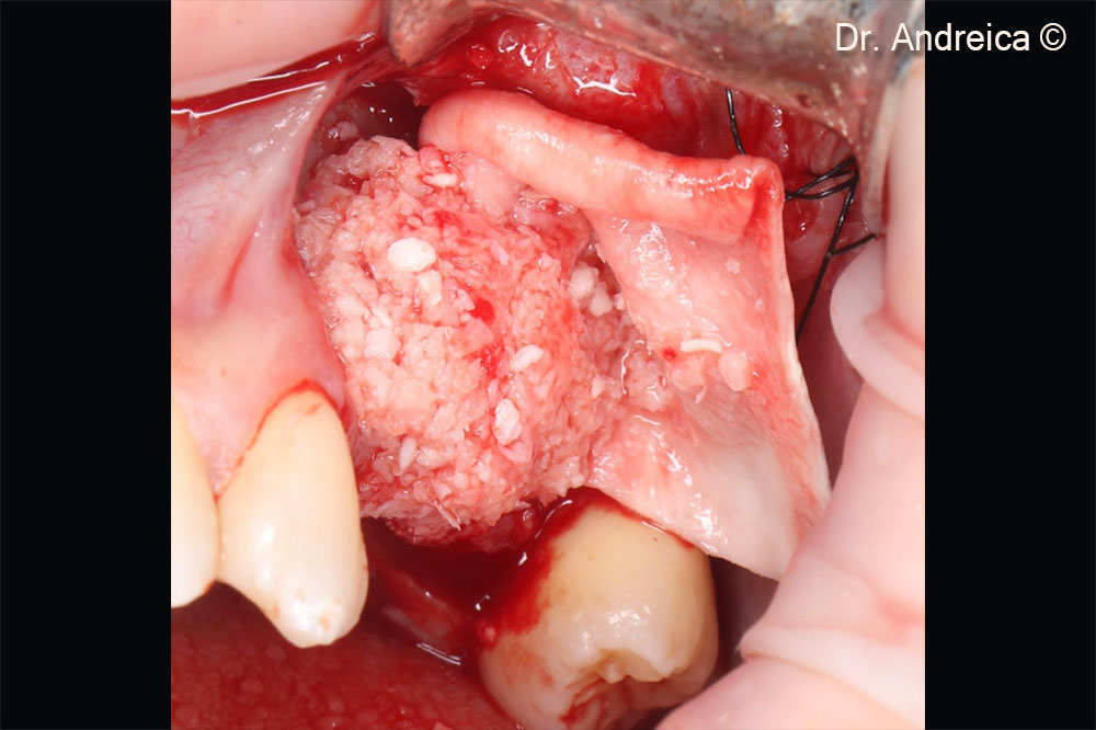
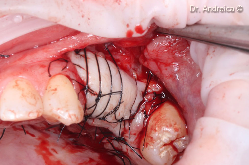
Used biomaterials
- Apatos®
- mp3®
- Gen-Os®
- Evolution
OsteoBiol by Tecnoss
Attention please! The OsteoBiol® website contains information on Medical Devices, which may be dangerous for the patient health and safety if not used exclusively by medical professionals.