Implant placement in the estethic area: the in-between implant
Dr. Eric Van Dooren
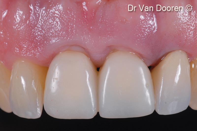
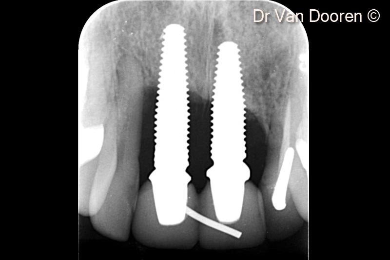
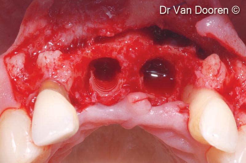
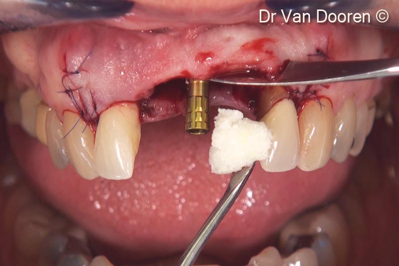
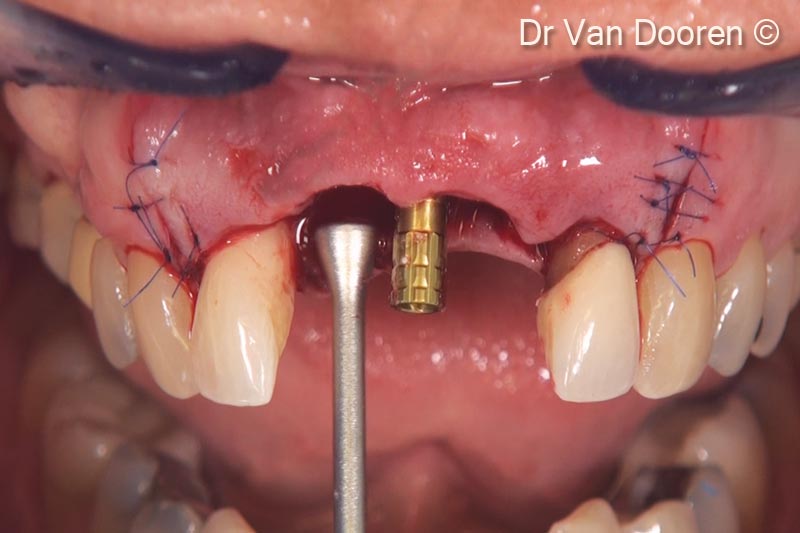
Initial situation
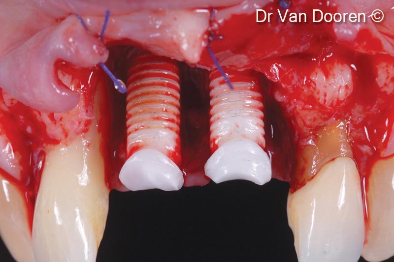
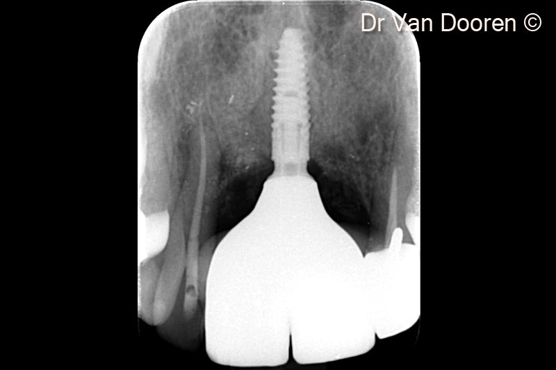
A female patient (46 years old) shows severe peri-implantitis around two implants
OsteoBiol by Tecnoss
Attention please! The OsteoBiol® website contains information on Medical Devices, which may be dangerous for the patient health and safety if not used exclusively by medical professionals.