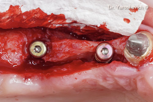
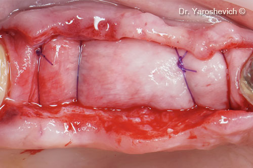
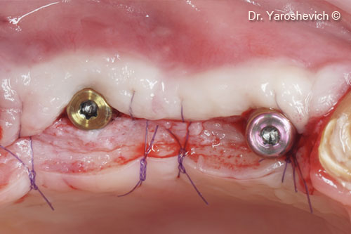
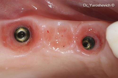
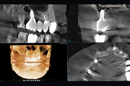
Horizontal bone augmentation with a collagenated xenograft
Dr. Pavel Yaroshevich
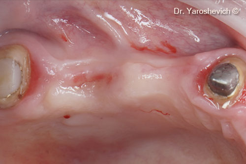
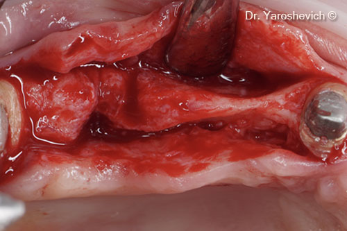
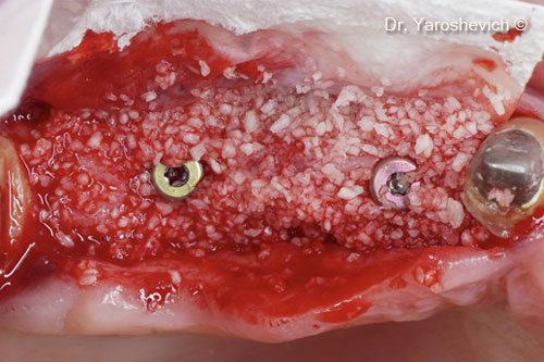
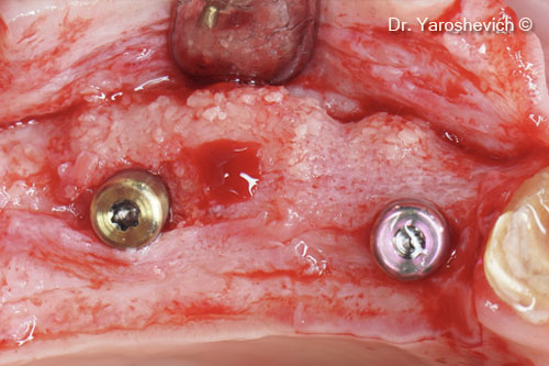
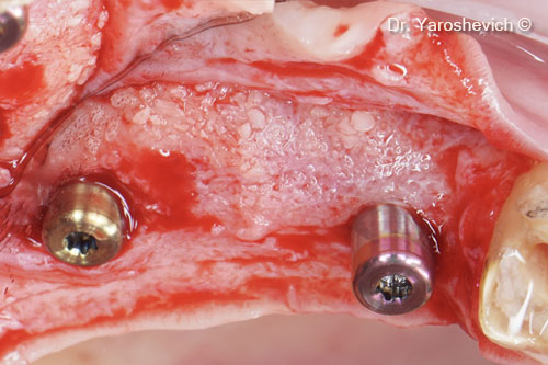
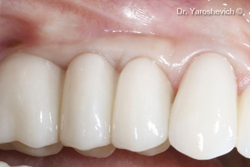
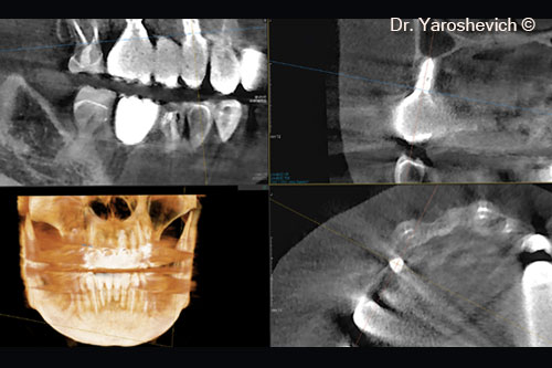
Initial situation
A female patient (45 years old) shows a knife-edge ridge
OsteoBiol by Tecnoss
Attention please! The OsteoBiol® website contains information on Medical Devices, which may be dangerous for the patient health and safety if not used exclusively by medical professionals.