Subcrestal minimal invasive sinus augmentation
Dr. Ziv Mazor
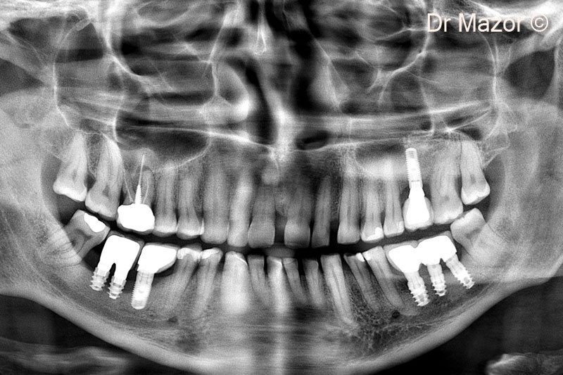
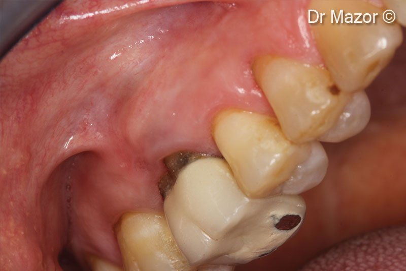
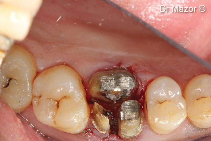
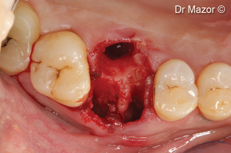
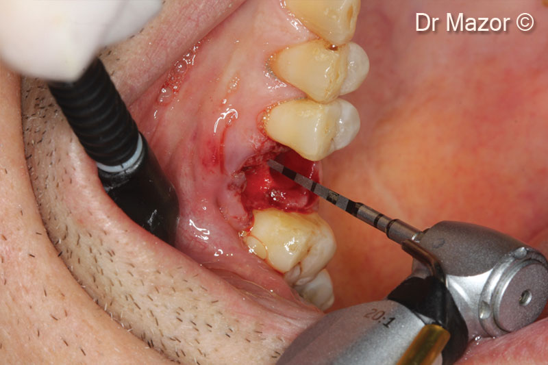
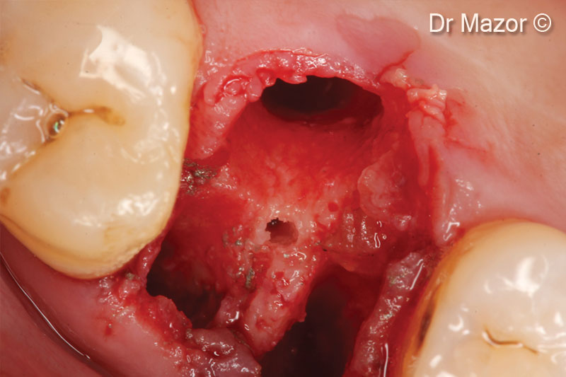
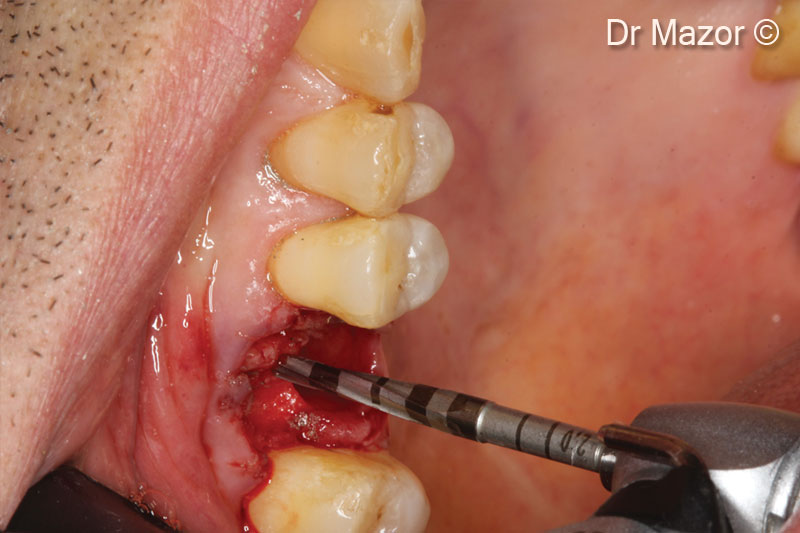
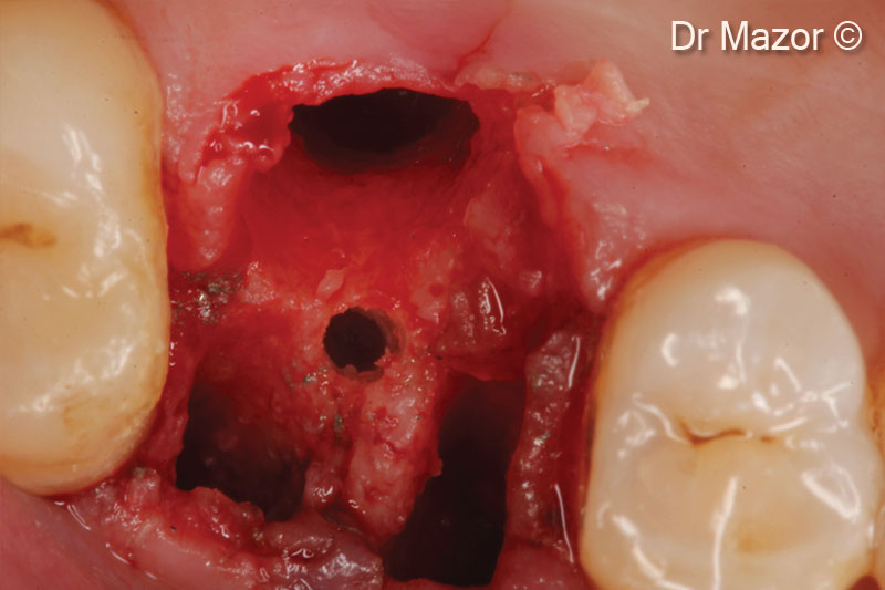
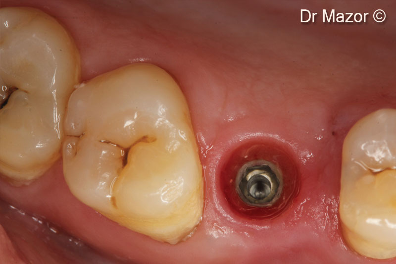
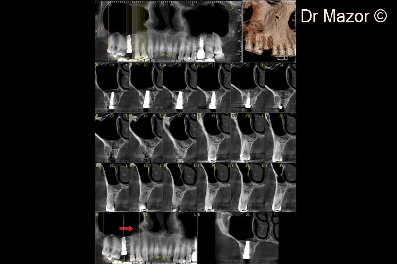
Initial situation
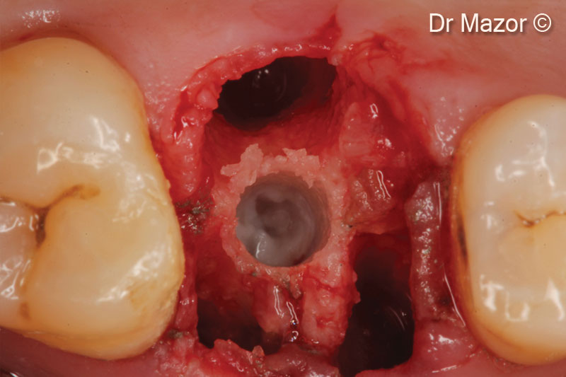
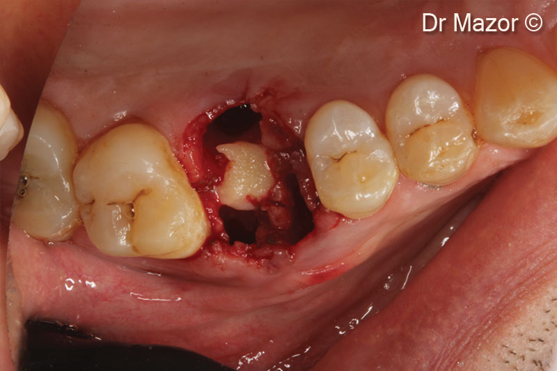
A male patient (49 years old) shows a hopeless tooth
OsteoBiol by Tecnoss
Attention please! The OsteoBiol® website contains information on Medical Devices, which may be dangerous for the patient health and safety if not used exclusively by medical professionals.