A male patient (35 years old) shows a vertical defect in the esthetic area
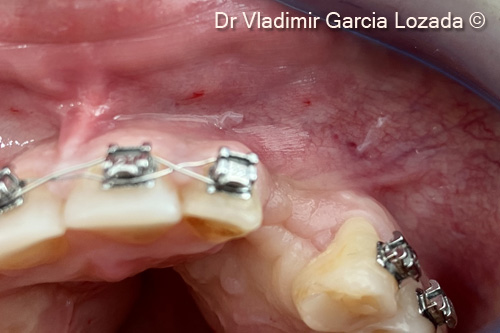
1. Initial clinical situation
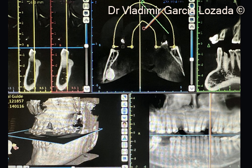
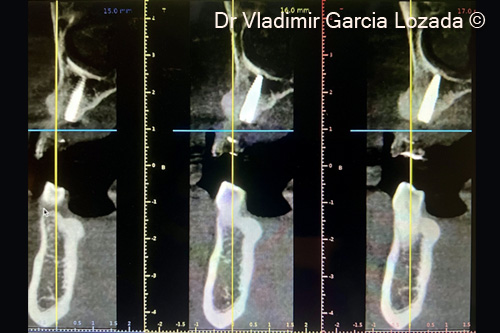
2. CBCT pre-op initial situation
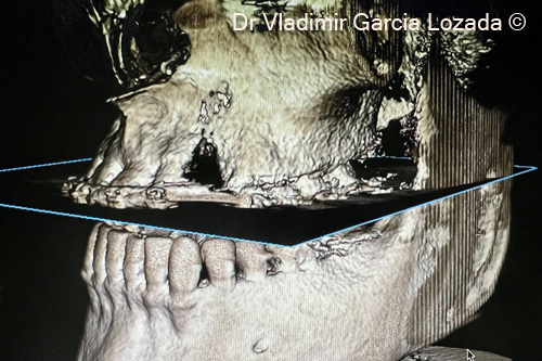
3. CBCT bone defect in site #23
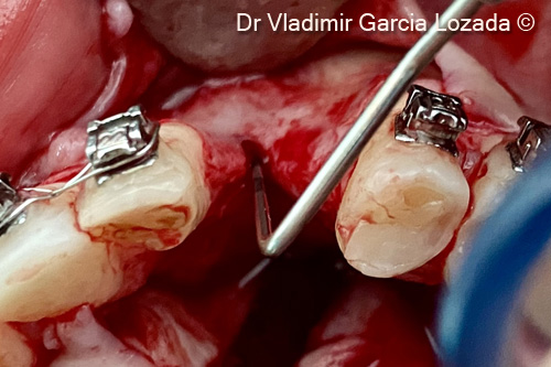
4. Degloving of a full thickness flap observing the three-dimensional defect in site #23
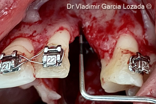
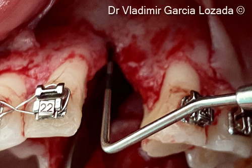
5. Measurement of the defect with a deep probe
6. Axial clinical view of the three-dimensional defect
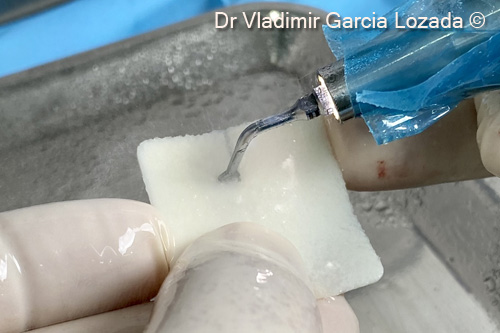
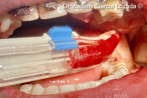
7. Preparation of the OsteoBiol® Soft Cortical Lamina® using a piezosurgery device
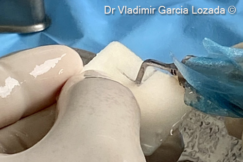
8. Preparation of the OsteoBiol® Soft Cortical Lamina® according to the size of the bone defect site
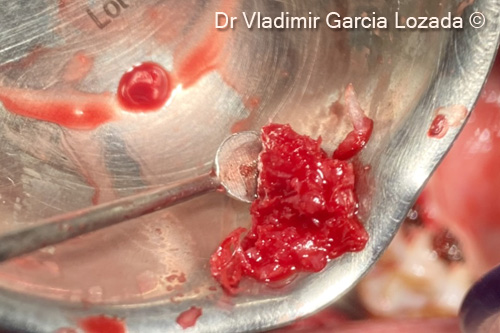
9. Recollection of autogenous bone chips with a safescraper from the mandibular ramus
10. Particulated autogenous bone chips recollected
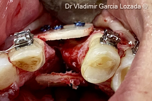
11. Buccal and palatal OsteoBiol® Soft Cortical Lamina® fixation with 4 ostheosyntesis screws
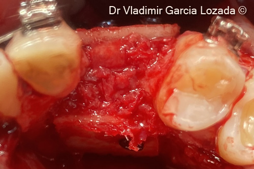
12. Bone defect filling inside the 2 OsteoBiol® Soft Cortical Laminas with autogenous bone chips
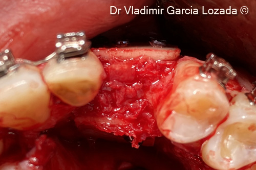
13. Axial clinical view of the bone augmentation before flap closure
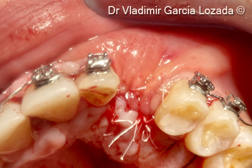
14. Management of soft tissues and flap closure using 5-0 PTFE suture
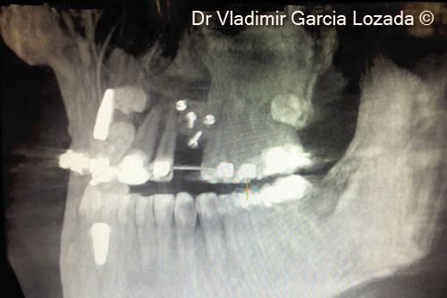
15. CBCT post-op, lateral view
16. CBCT post-op, axial view
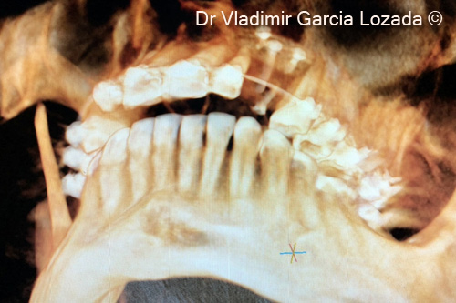
17. CBCT 9 month after implant placement in site #23 with relining procedure, recommended in these cases
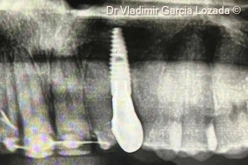
18. X-ray with final restoration
Attention please! The OsteoBiol® website contains information on Medical Devices, which may be dangerous for the patient health and safety if not used exclusively by medical professionals.