Treatment of a complex defect in the estetich area

Dr. Abdelsalam Elaskary
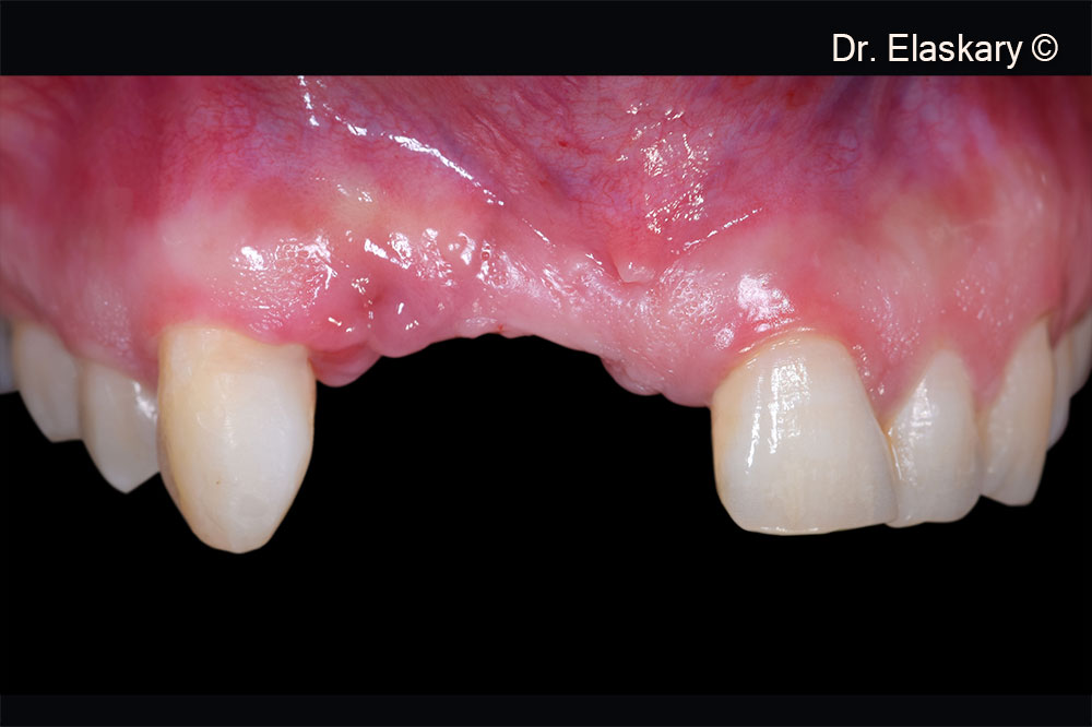
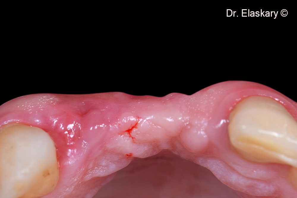
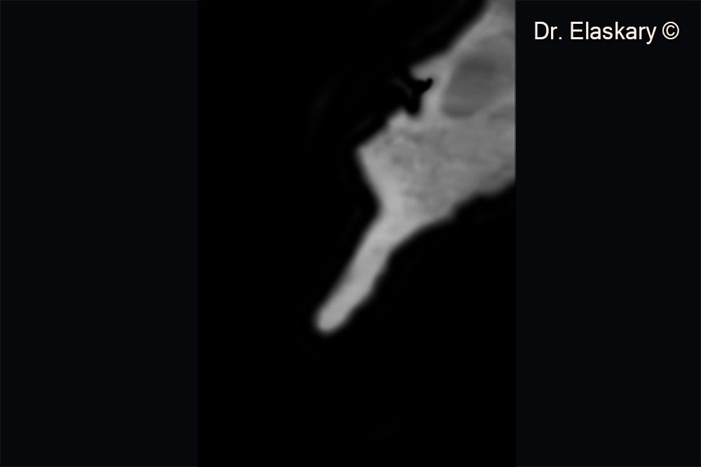
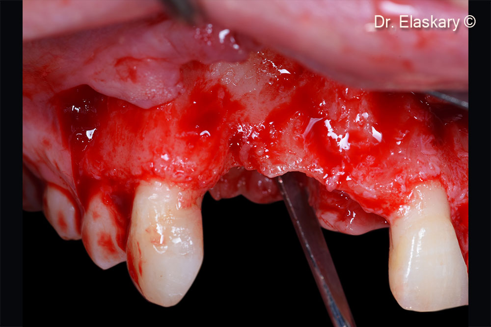
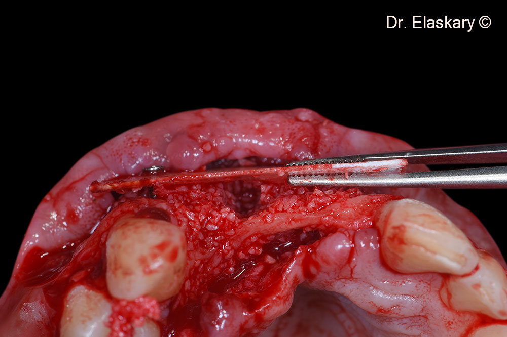
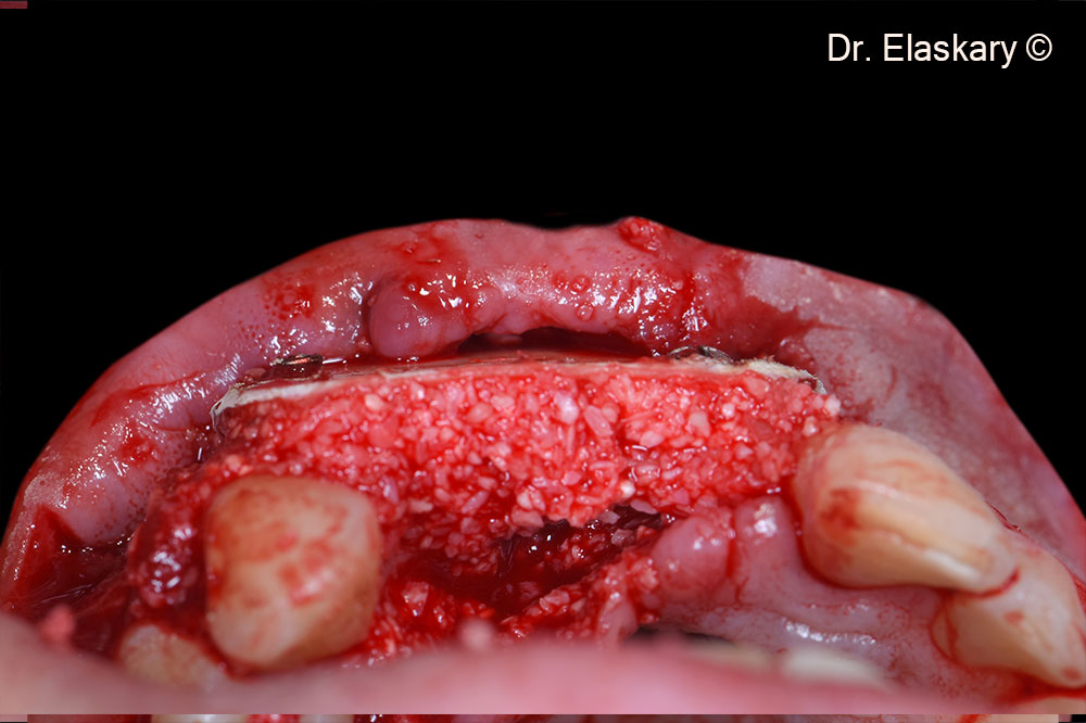
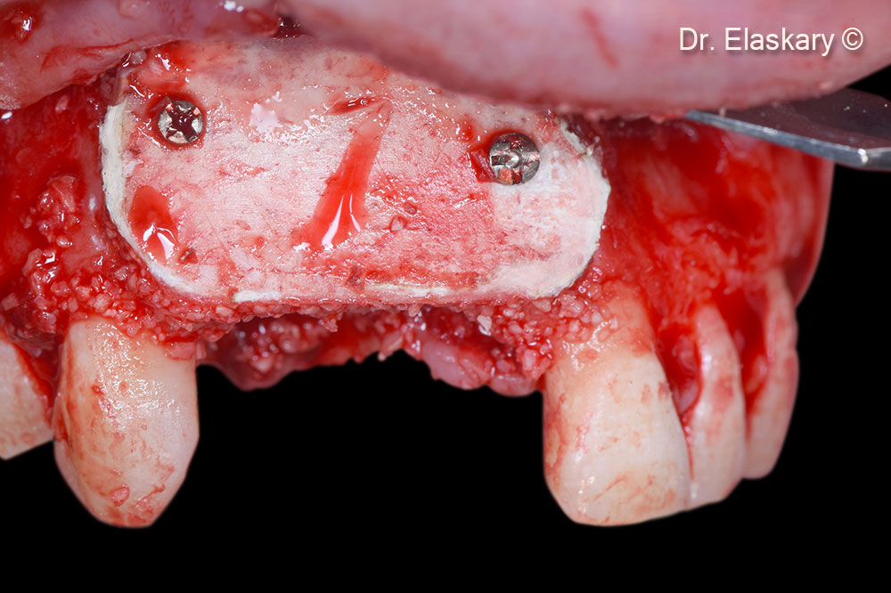
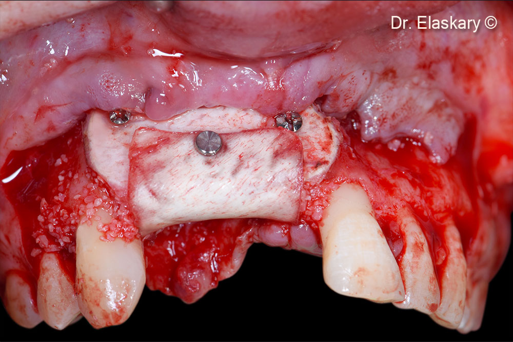
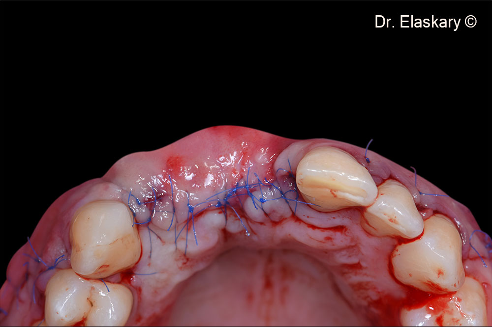
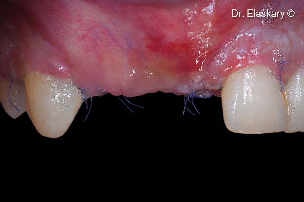
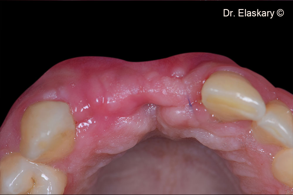
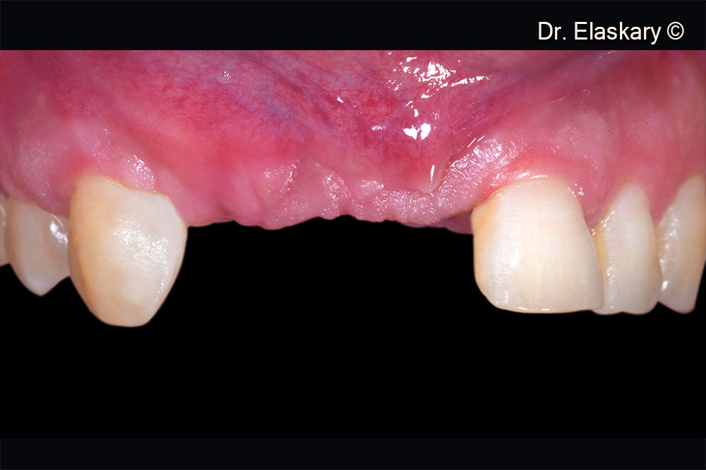
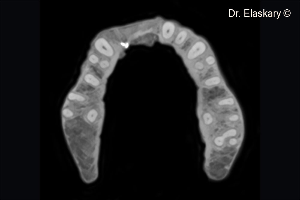
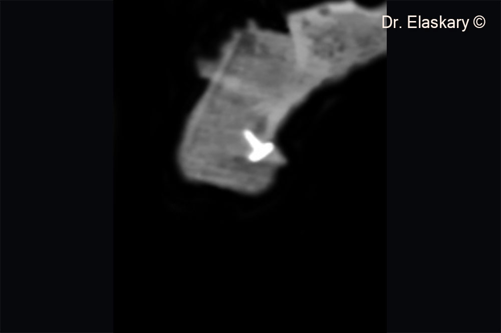
Initial situation

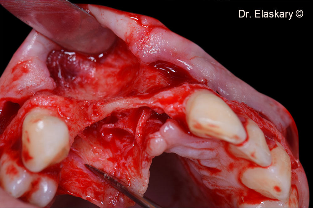
In a female patient (51 years old) the buccal bone is missing

Used biomaterials
  • Gen-Os®
  • Lamina®
Pre operative frontal view of lost both vertical and horizontal anterior tissues related to the alveolar ridge

Pre operative frontal view of lost both vertical and horizontal anterior tissues related to the alveolar ridge

Pre operative incisal view of lost both vertical and horizontal anterior tissues related to the alveolar ridge

Pre operative incisal view of lost both vertical and horizontal anterior tissues related to the alveolar ridge

CBCT scan showing the loss of the buccal bone

CBCT scan showing the loss of the buccal bone

Intra surgical incisal view showing the total loss of the osseous foundation related to the missing teeth

Intra surgical incisal view showing the total loss of the osseous foundation related to the missing teeth

Intra surgical incisal view showing the total loss of the osseous foundation related to the missing teeth

Intra surgical incisal view showing the total loss of the osseous foundation related to the missing teeth

Lamina® was stabilized on top of the defect leaving the regenerative space underneath Gen-Os®

Lamina® was stabilized on top of the defect leaving the regenerative space underneath Gen-Os®

The regenerative space was filled with Gen-Os® mixed with autogenous cortical chips.

The regenerative space was filled with Gen-Os® mixed with autogenous cortical chips.

Lamina® is stabilized from the palatal to the labial side to protect the bone graft occlusally

Lamina® is stabilized from the palatal to the labial side to protect the bone graft occlusally

Lamina® is stabilized from the palatal to the labial side to protect the bone graft occlusally

Lamina® is stabilized from the palatal to the labial side to protect the bone graft occlusally

Lamina® protect the bone graft occlusally

Lamina® protect the bone graft occlusally

Frontal view showing the flap released, extended and closed using 6-0 proline sutures.

Frontal view showing the flap released, extended and closed using 6-0 proline sutures.

Incisal view showing 3 weeks post surgery showing a total tissue closure.

Incisal view showing 3 weeks post surgery showing a total tissue closure.

Frontal view showing 3 weeks post surgery showing a total tissue closure.

Frontal view showing 3 weeks post surgery showing a total tissue closure.

CBCT scan showing the total regeneration of the lost tissues.

CBCT scan showing the total regeneration of the lost tissues.

CBCT scan showing the total regeneration of the lost tissues.

CBCT scan showing the total regeneration of the lost tissues.

Used biomaterials
genos
Gen-Os®
lamina
Lamina®
Are you a medical professional?
No

Attention please! The OsteoBiol® website contains information on Medical Devices, which may be dangerous for the patient health and safety if not used exclusively by medical professionals.