Treatment of an atrophic ridge

Prof. Antonio Barone
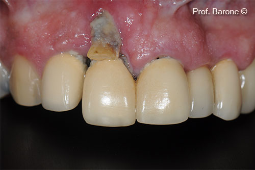
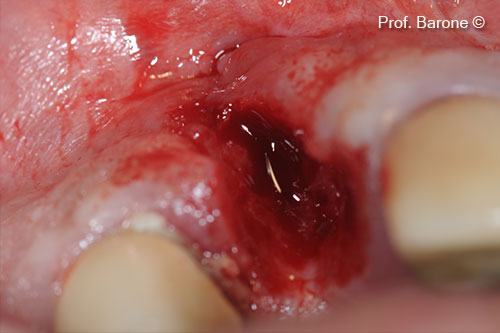
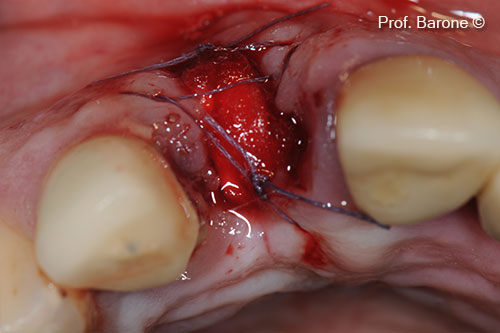
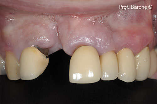
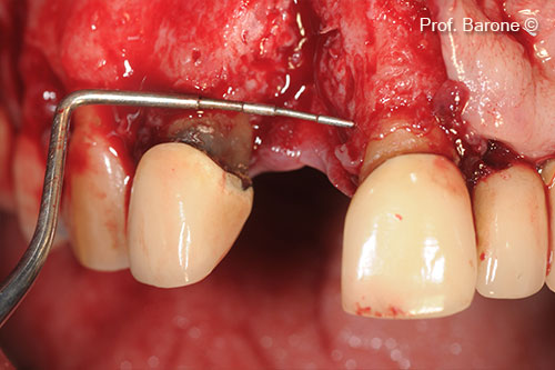
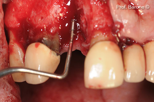
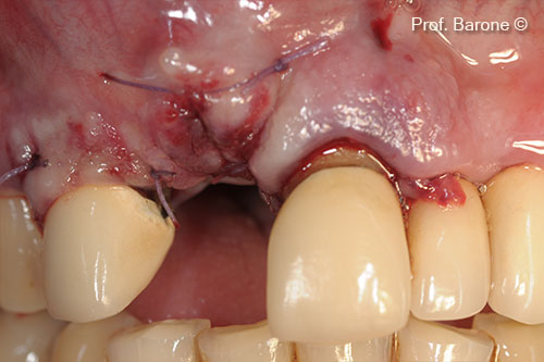
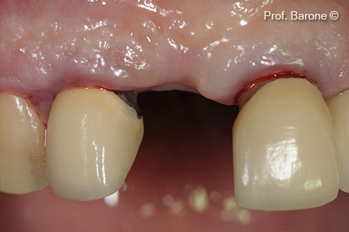
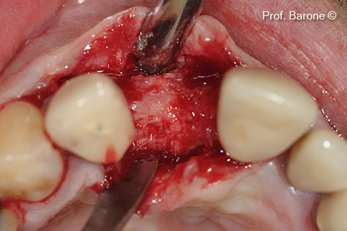
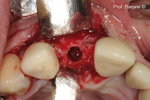
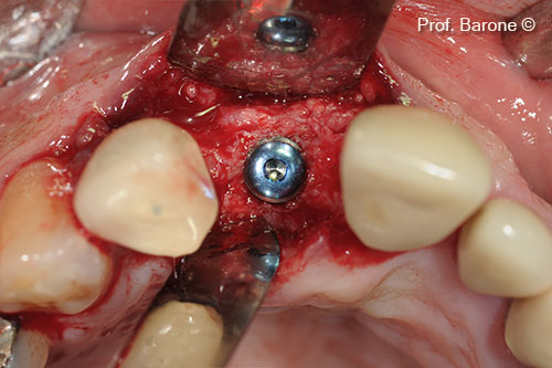
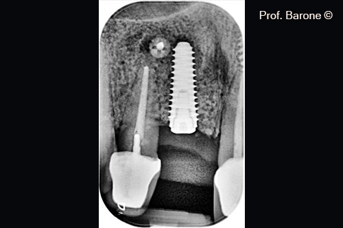
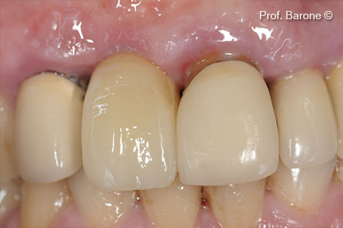
Initial situation

A male patient (47 years old) shows an esthetic imperfection in position 11
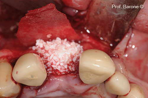
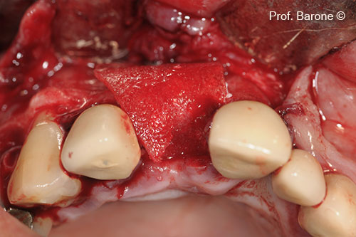
Used biomaterials
  • GTO®
  • Lamina®
Buccal View, tooth #11

Buccal View, tooth #11

Tooth Extraction

Tooth Extraction

Fresh Extraction socket treated with collagen sponge and cross-suture

Fresh Extraction socket treated with collagen sponge and cross-suture

Healing after 3 months

Healing after 3 months

Residual Bone Defect, mesio-distal dimension

Residual Bone Defect, mesio-distal dimension

: Residual Bone Defect, vertical dimension

: Residual Bone Defect, vertical dimension

Bone Defect grafted with GTO®

Bone Defect grafted with GTO®

Lamina® Soft to protect the graft

Lamina® Soft to protect the graft

Soft tissue primary closure

Soft tissue primary closure

Soft Tissue Healing after 6 months

Soft Tissue Healing after 6 months

Augmented Ridge

Augmented Ridge

Implant site preparation

Implant site preparation

Implant inserted in the augmented ridge

Implant inserted in the augmented ridge

Periapical Radiograph 3 months after implant placement

Periapical Radiograph 3 months after implant placement

Final Crown

Final Crown

Used biomaterials
gto
GTO®
lamina
Lamina®
Are you a medical professional?
No

Attention please! The OsteoBiol® website contains information on Medical Devices, which may be dangerous for the patient health and safety if not used exclusively by medical professionals.