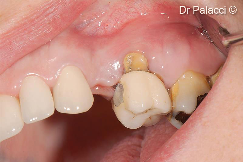
A female patient (54 years old) lost teeth 24,26,27 and 28
Used biomaterials
GTO®
1. Loss of #24, #26, #27 and #28, have to be extracted
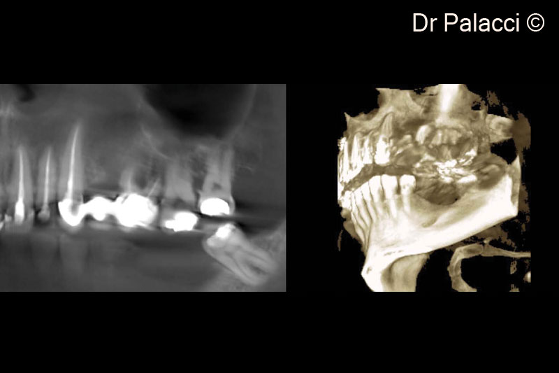
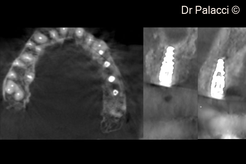
2. 3D X-rays, major bone loss around #26, #27, #28 furcation involvement. Significant concavity in #24
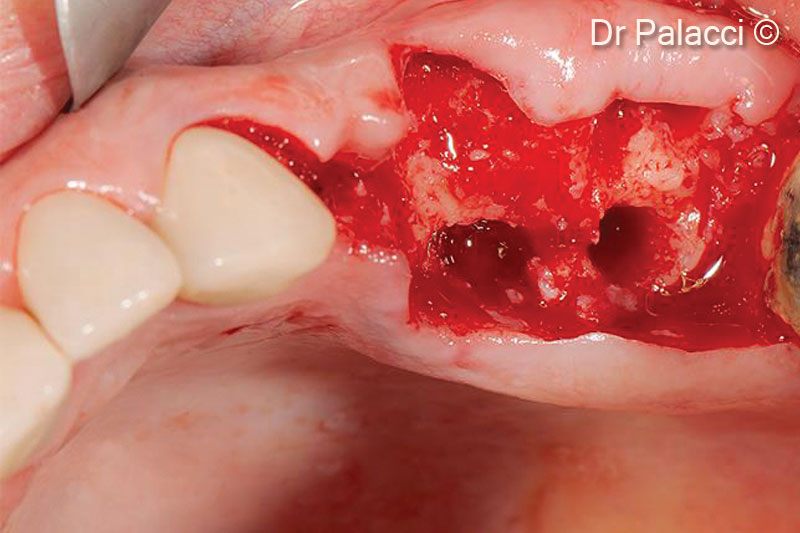
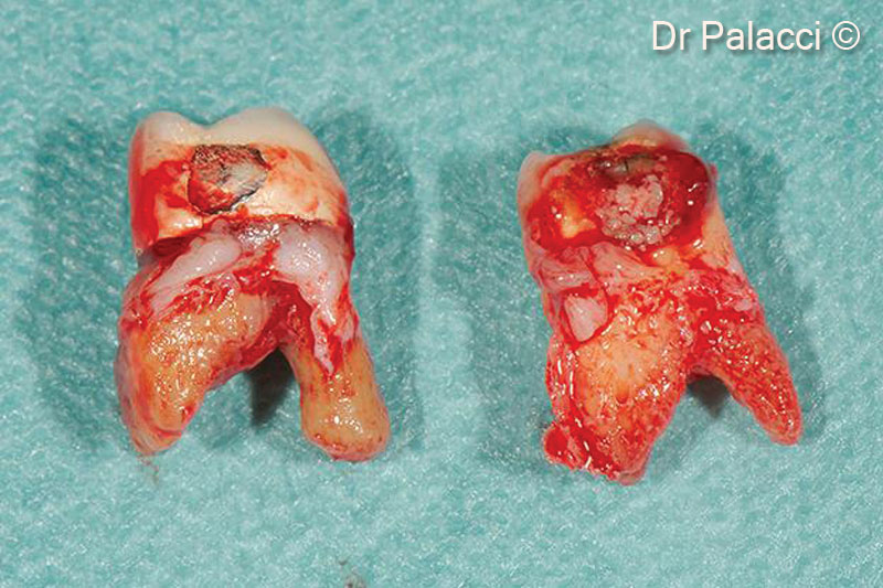
3. Teeth are extracted, all labial bone is lost. Major bone loss is visible
4. Teeth are extracted, all labial bone is lost. Major bone loss is visible
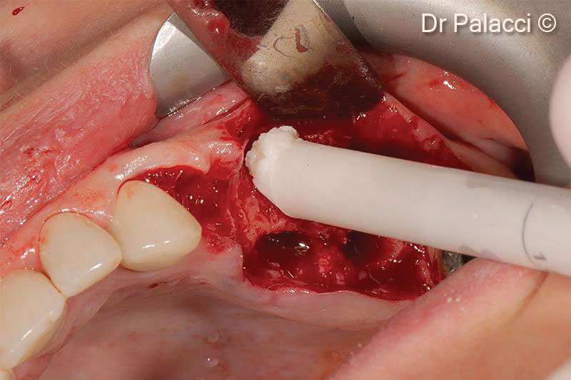
5. OsteoBiol® GTO® is inserted into the alveoli
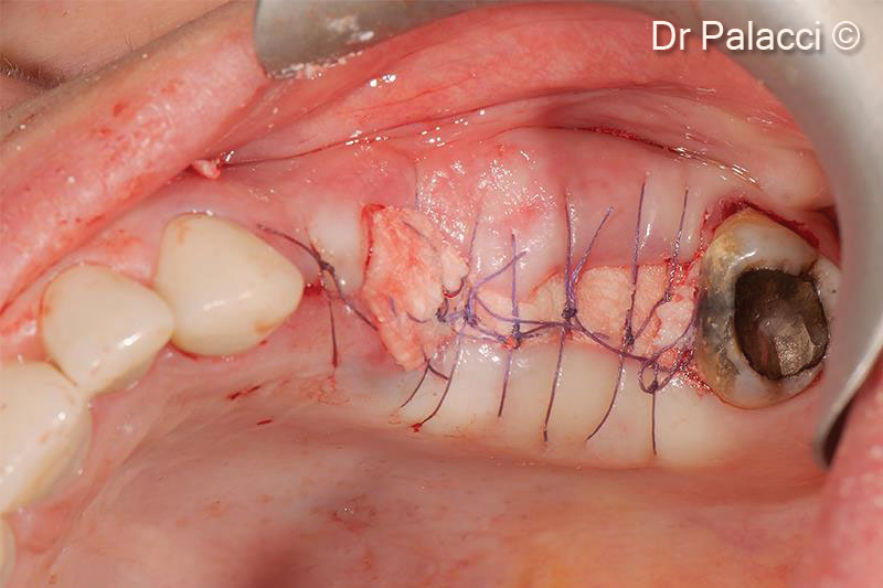
6. A collagen membrane covers the defect filled with the biomaterial
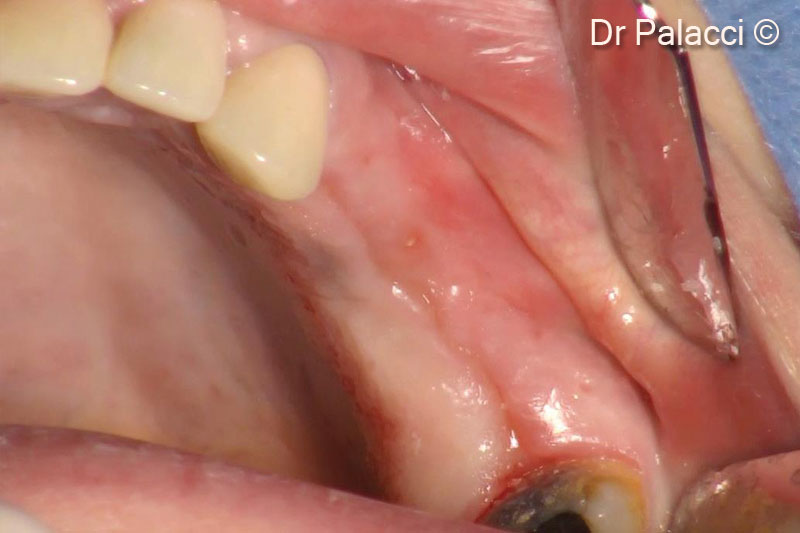
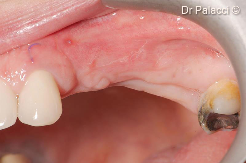
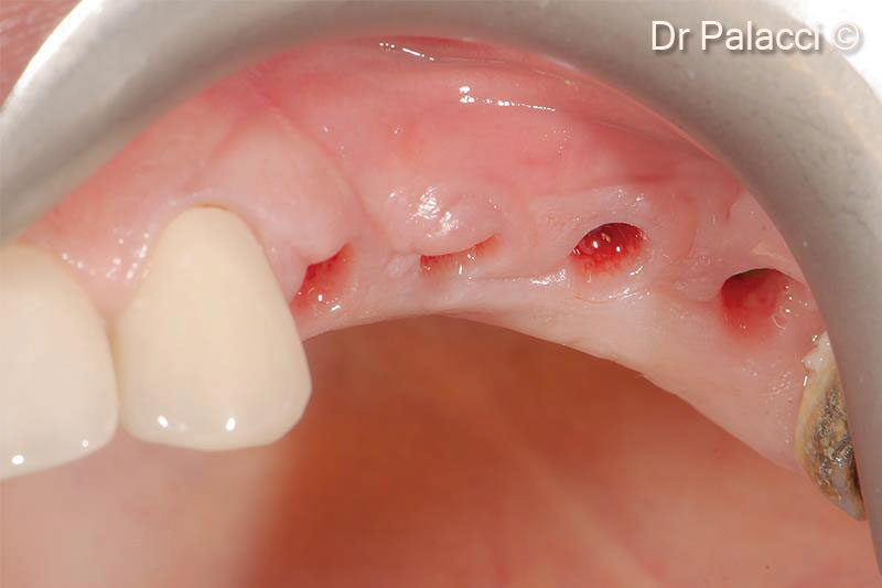
7. Re-entry 4 months later. Note the significant labial ridge augmentation as well as the soft tissue quality
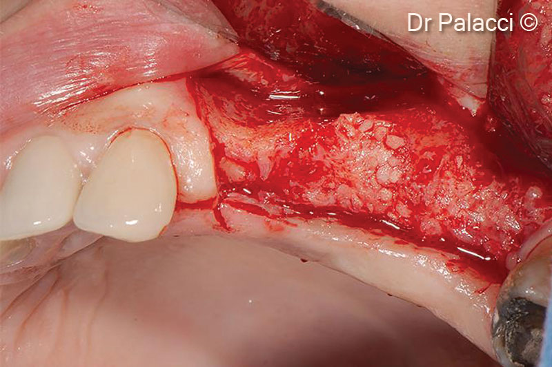
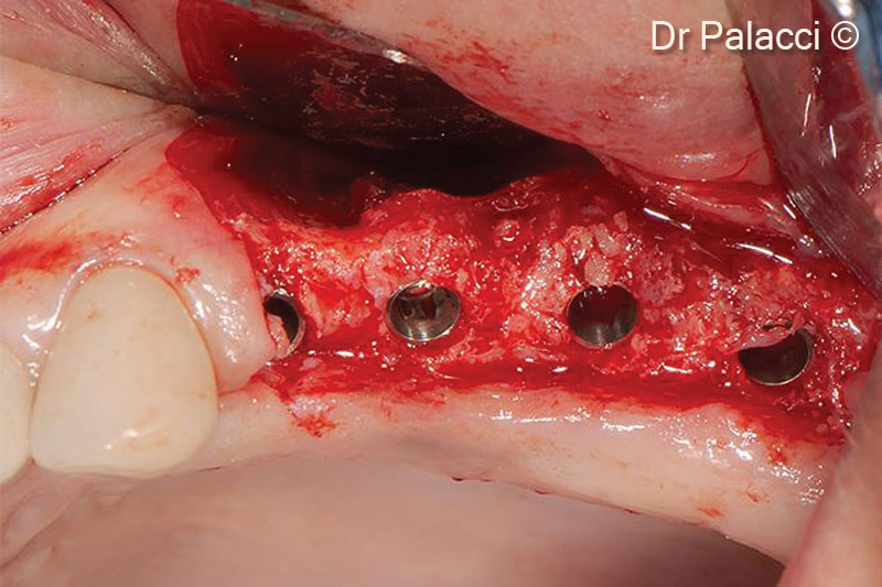
8. Note the optimal hard tissue volume obtained and the quality of the bone formation
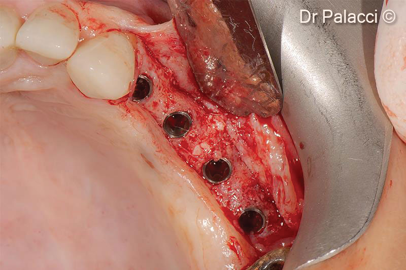
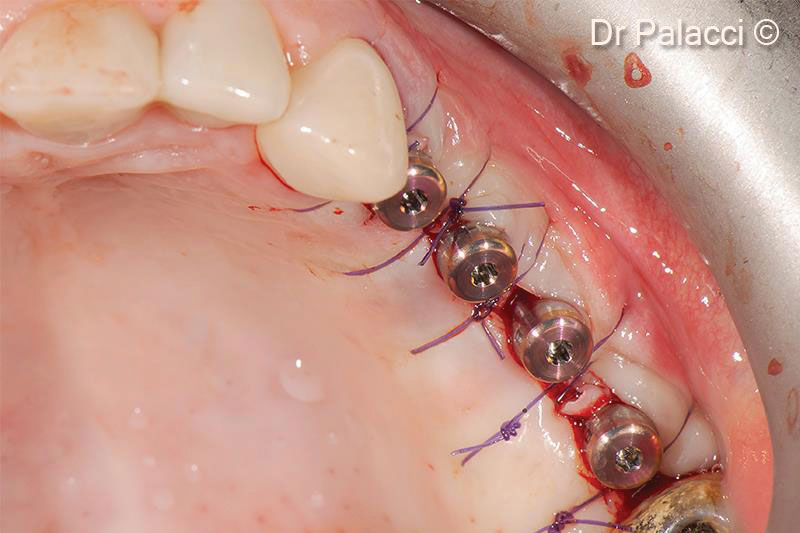
9. 4 implants of 3.5-mm diameter are inserted
10. X-ray illustrating the labial ridge augmentation
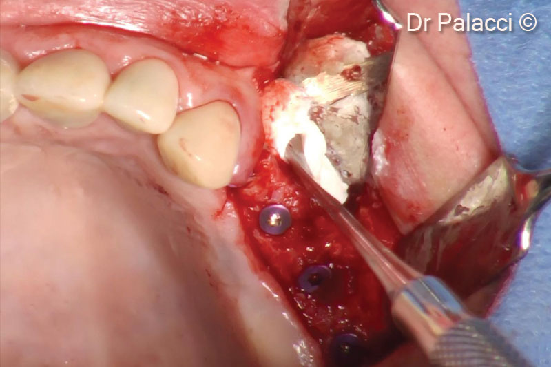
11. The concavity level in the apical portion of the first premolar, is filled with OsteoBiol® GTO® in order to recreate a natural ridge profile
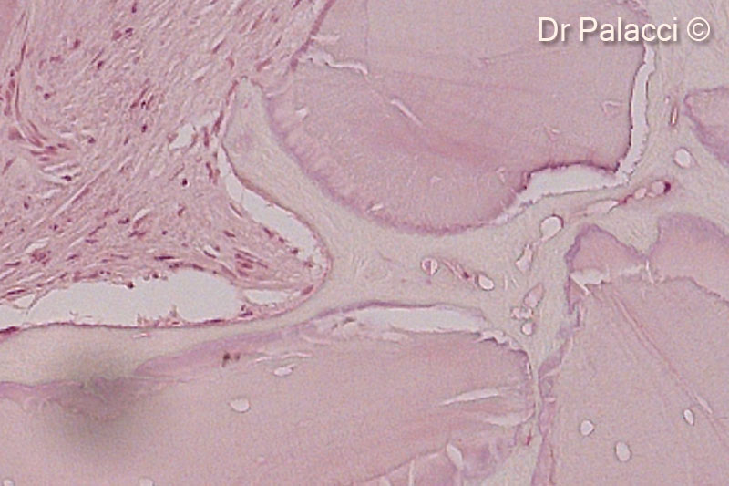
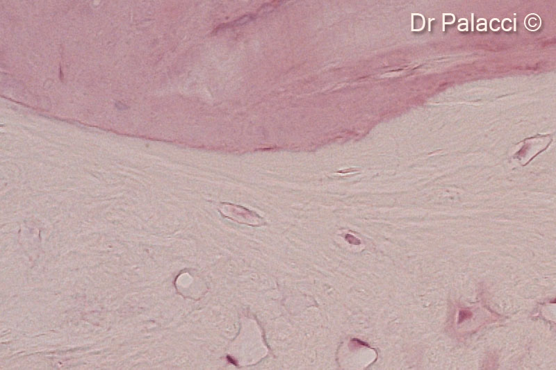
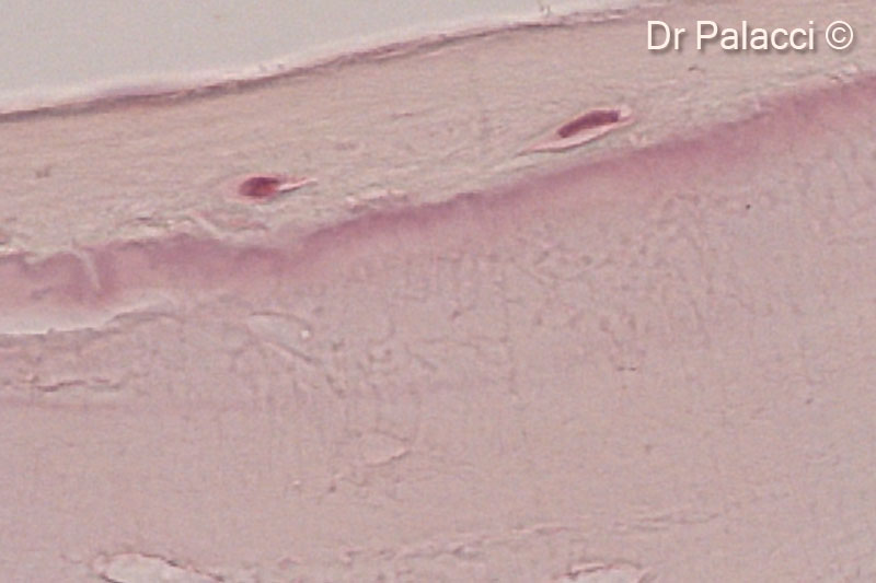
12. Part of biopsies retrieved 4 months after augmentation with GTO®. The particles of biomaterial are surrounded by newly formed bone, presence of osteocyte lacunae
13. Part of biopsies retrieved 4 months after augmentation with GTO®. The particles of biomaterial are surrounded by newly formed bone, presence of osteocyte lacunae
14. Part of biopsies retrieved 4 months after augmentation with GTO®. The particles of biomaterial are surrounded by newly formed bone, presence of osteocyte lacunae
15. 1 week post-op. Note the soft tissue healing
16. Re-entry 4 months later. Implants are perfectly integrated into a well vascularized bone
17. Soft tissue manipulation around implants
18. Healing after 3 weeks. Note the ridge augmentation as well as shape, colour, texture of soft tissue
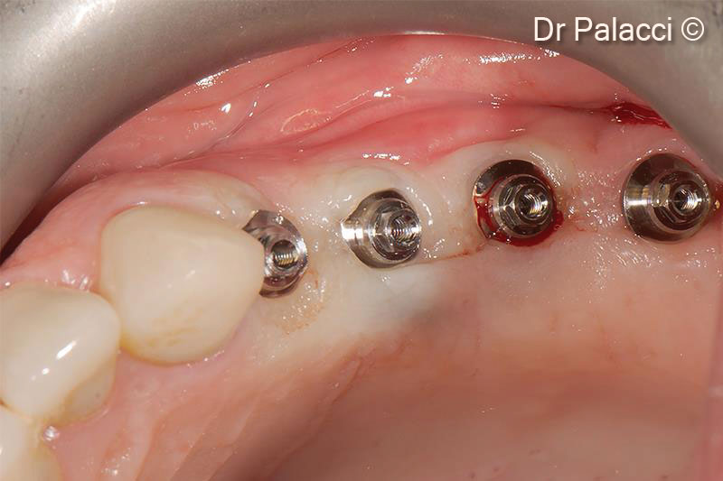
19. Abutments are selected and placed for a screw retained restauration
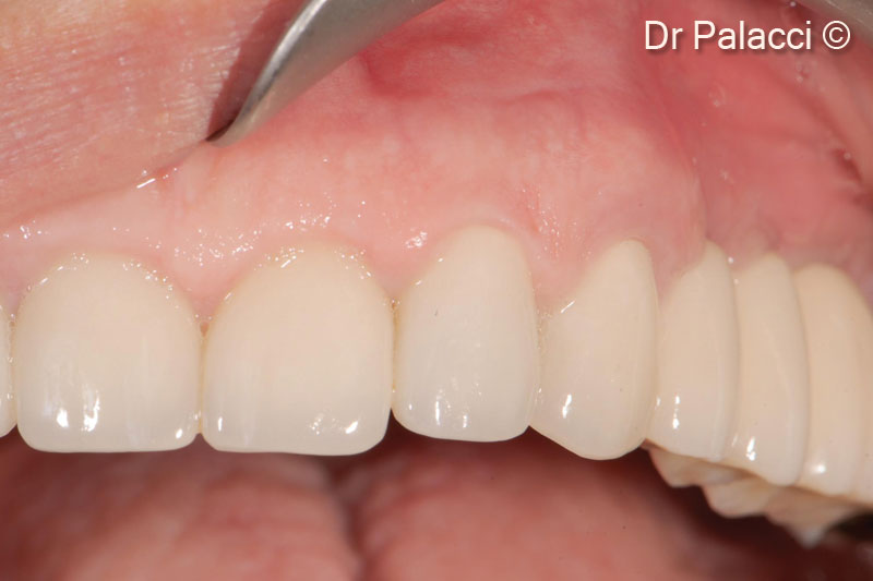
20. Final reconstruction in place (Prosthodontist Dr Christian Roux, Marseille)
Attention please! The OsteoBiol® website contains information on Medical Devices, which may be dangerous for the patient health and safety if not used exclusively by medical professionals.