Transcrestal sinus floor elevation with delayed implant insertion
Prof. Claudio Stacchi
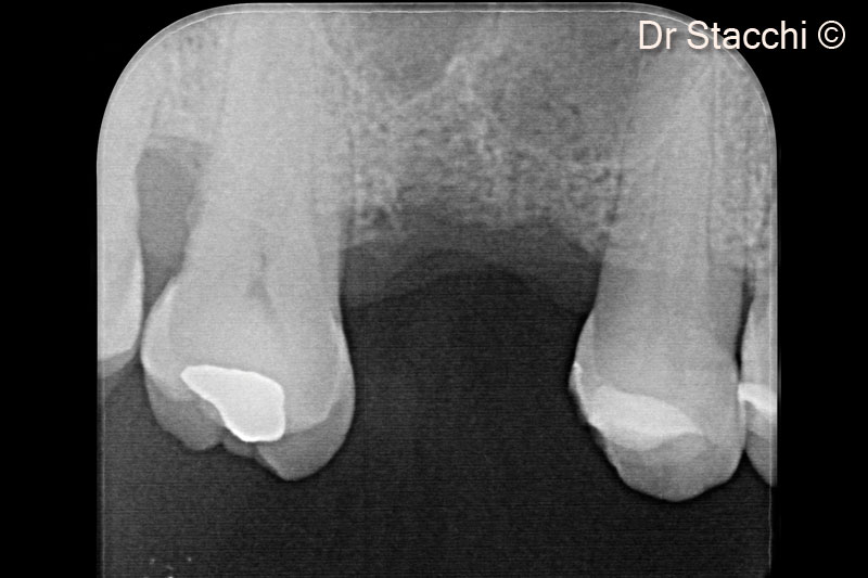
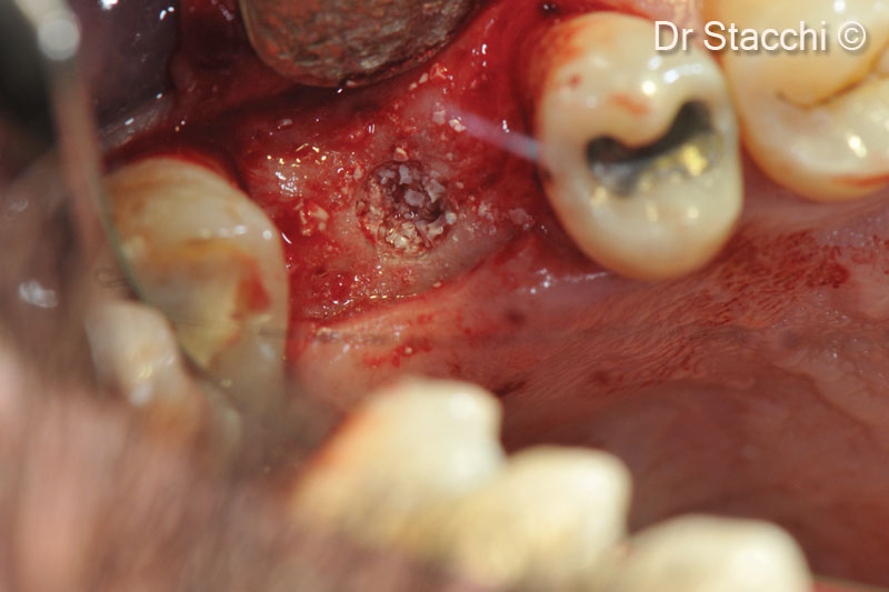
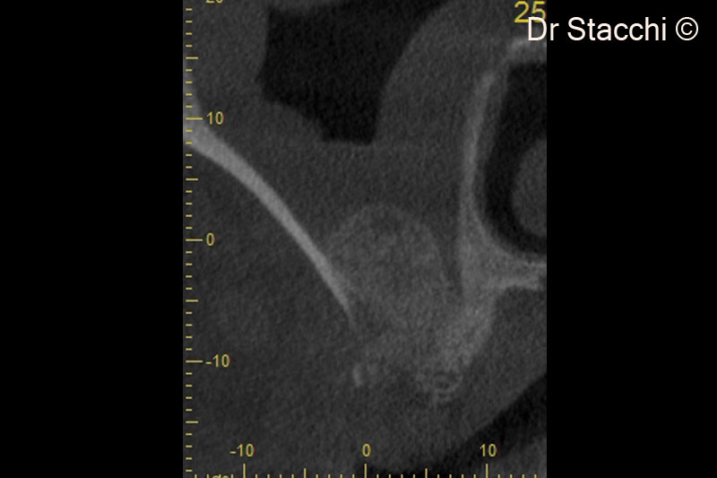
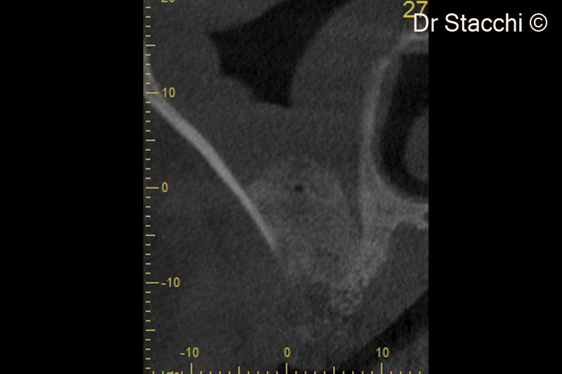
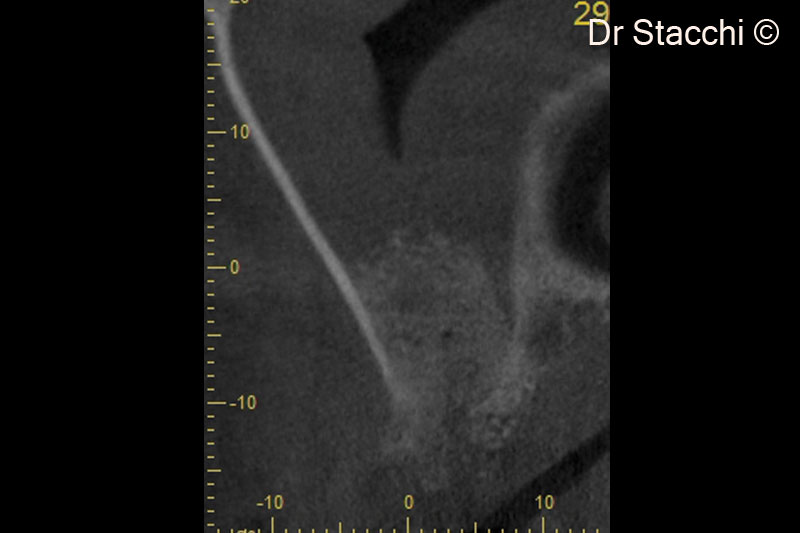
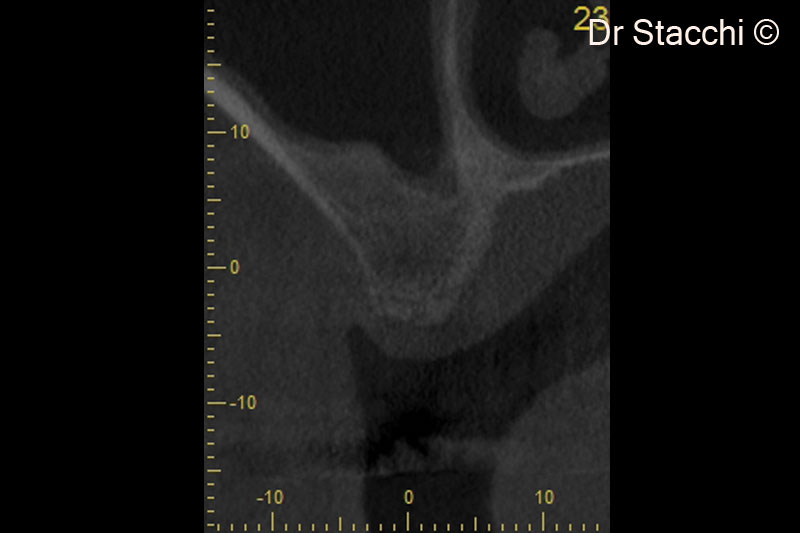
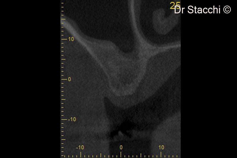
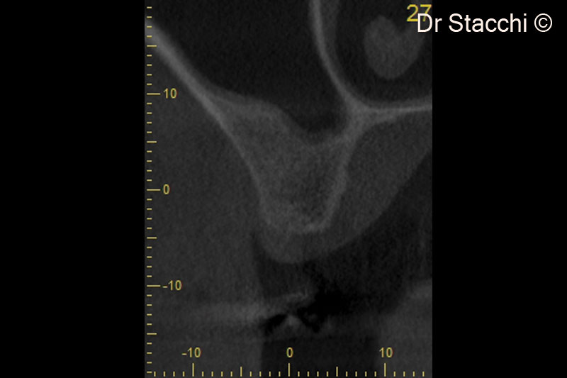
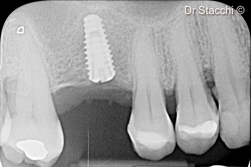
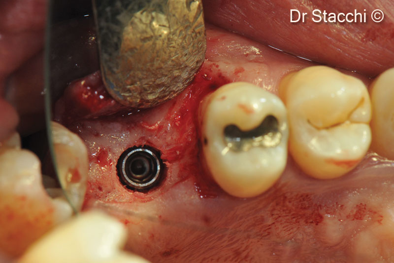
Initial situation
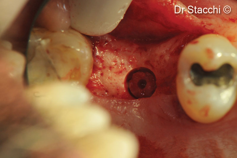
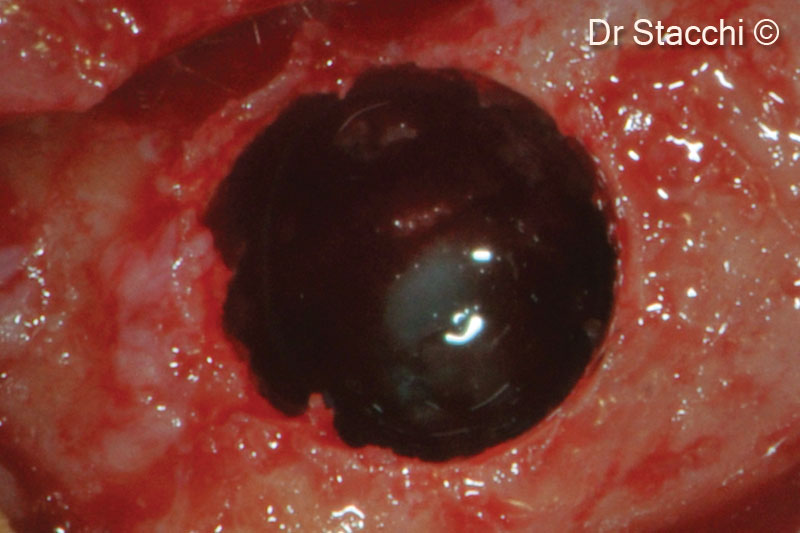
Based on the residual bone height, a female patient (66 years old) is considered eligible for transcrestal sinus floor elevation with delayed implant placement
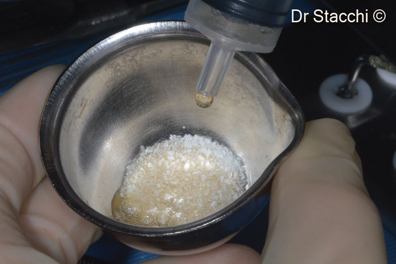
OsteoBiol by Tecnoss
Attention please! The OsteoBiol® website contains information on Medical Devices, which may be dangerous for the patient health and safety if not used exclusively by medical professionals.