Soft and hard tissues preservation in a compromised alveolar socket
Prof. Antonio Barone
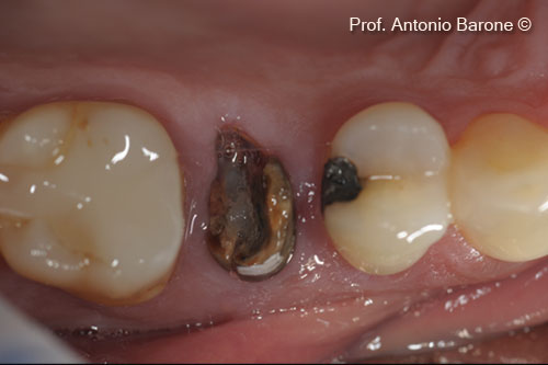
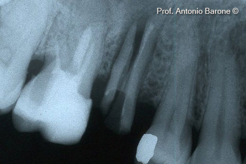
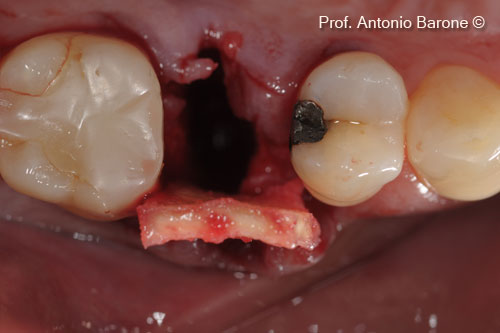
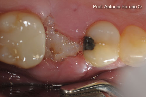
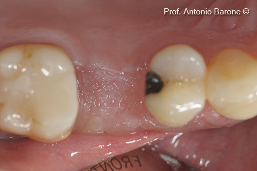
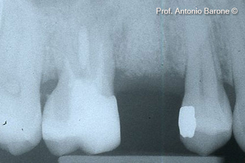
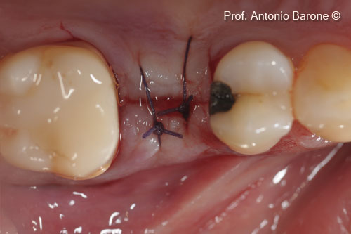
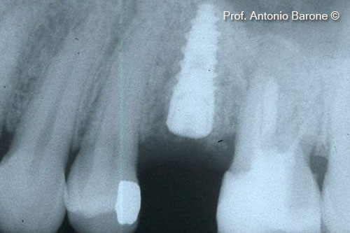
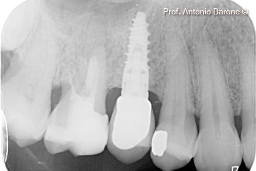
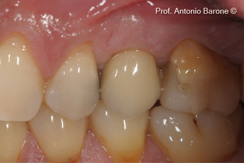
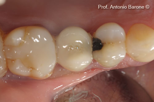
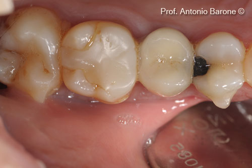
Initial situation
A female patient (54 years old) shows a fractured tooth in position 15
OsteoBiol by Tecnoss
Attention please! The OsteoBiol® website contains information on Medical Devices, which may be dangerous for the patient health and safety if not used exclusively by medical professionals.