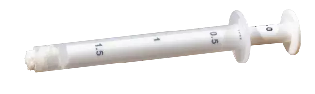
Socket rebuilding for immediate implant placement
Dr. Gerd Körner
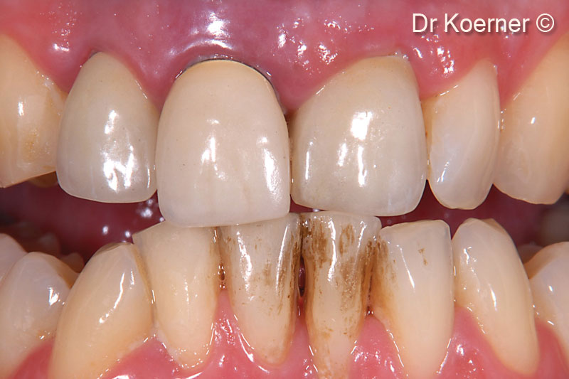
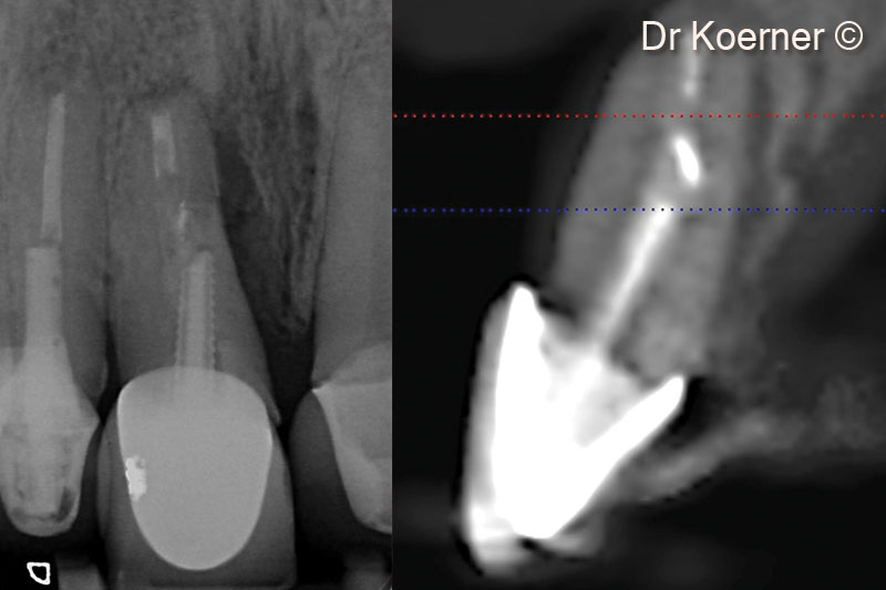
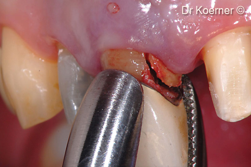
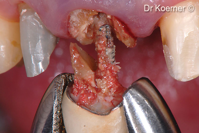
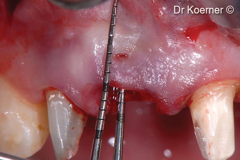
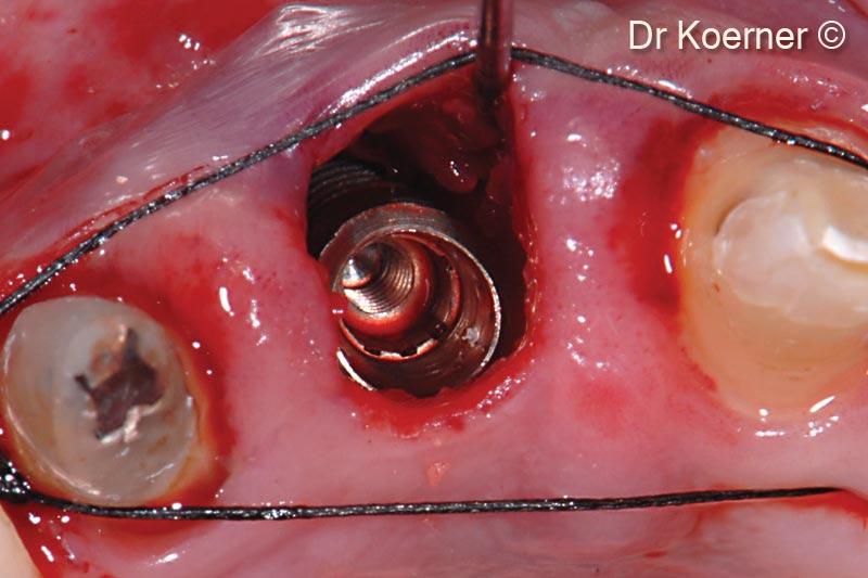
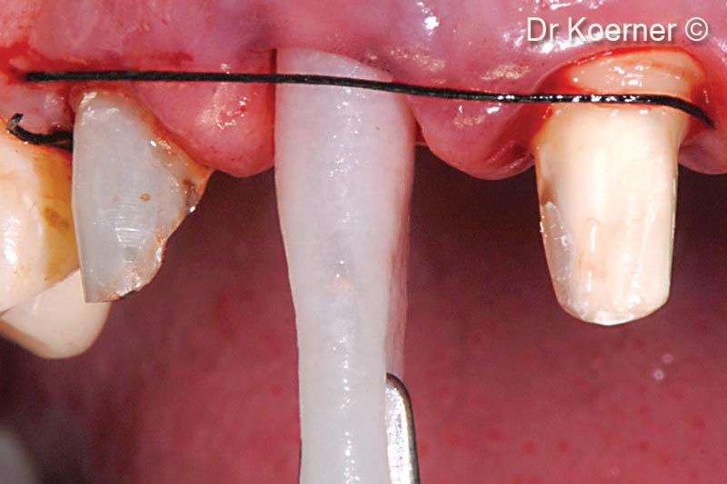
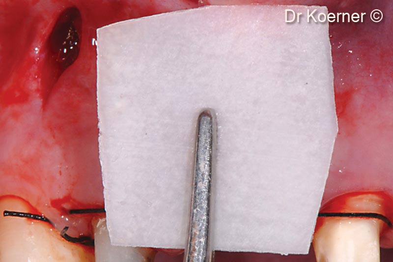
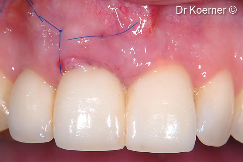
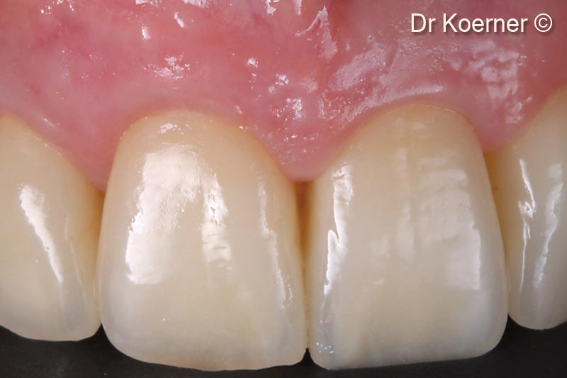
Initial situation
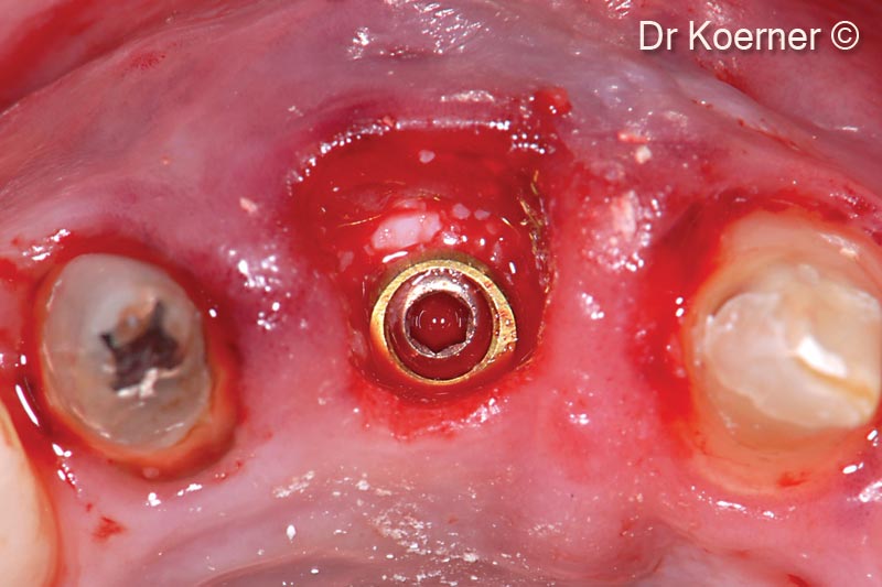
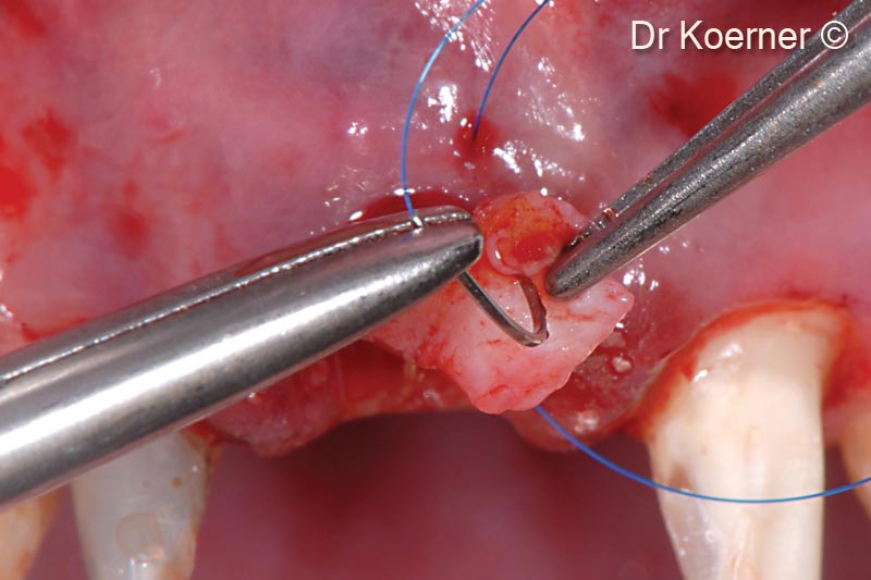
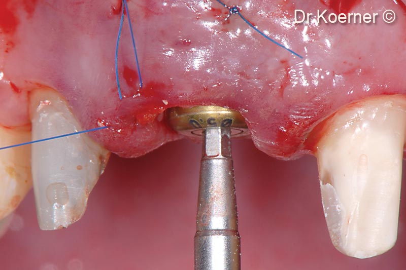
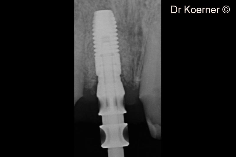
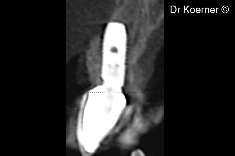
A female patient (45 years old) shows a hopeless tooth with missing buccal bone
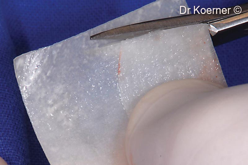
OsteoBiol by Tecnoss
Attention please! The OsteoBiol® website contains information on Medical Devices, which may be dangerous for the patient health and safety if not used exclusively by medical professionals.