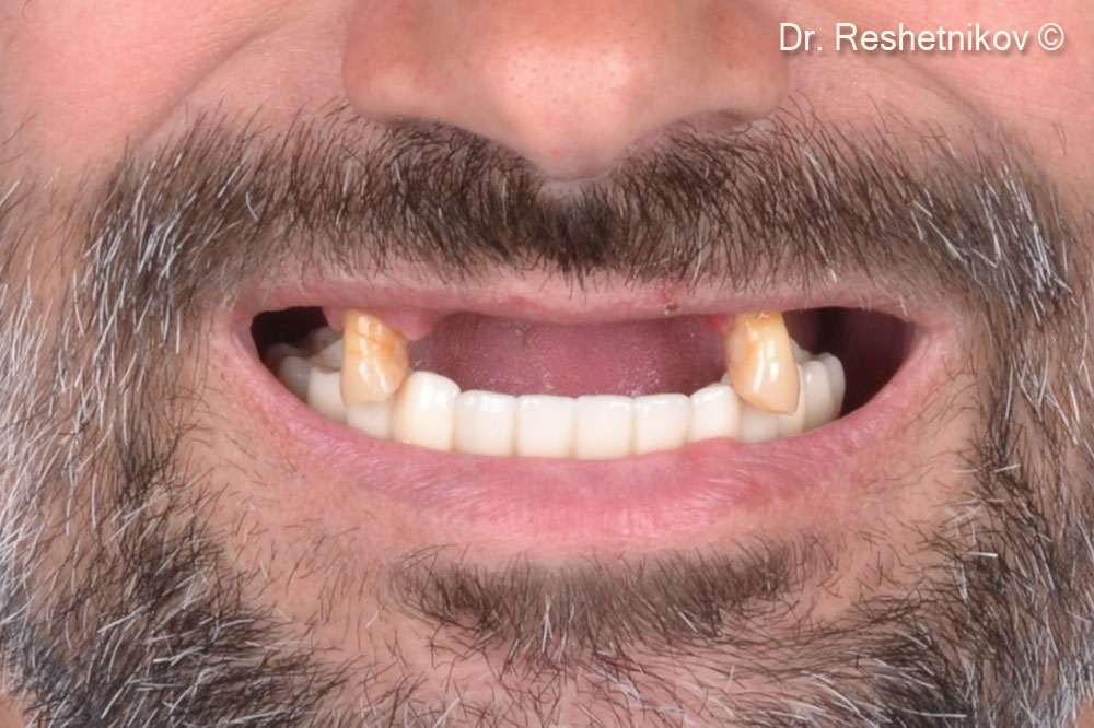
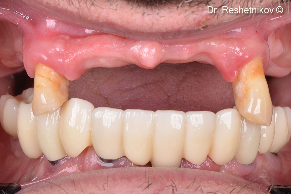
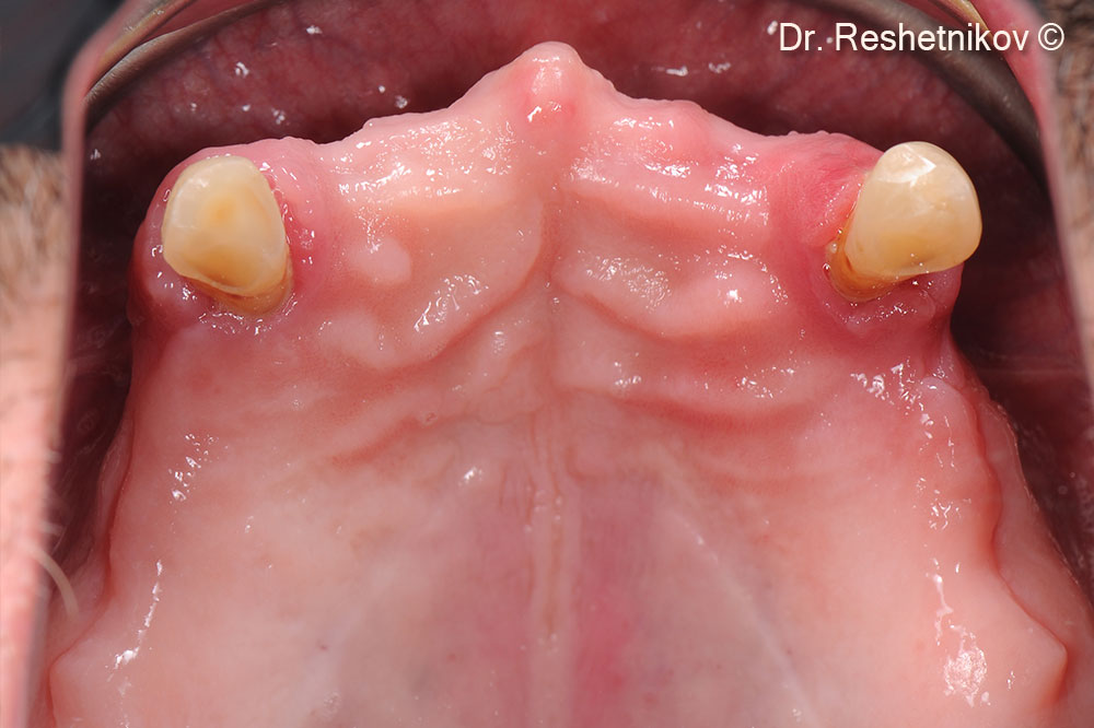
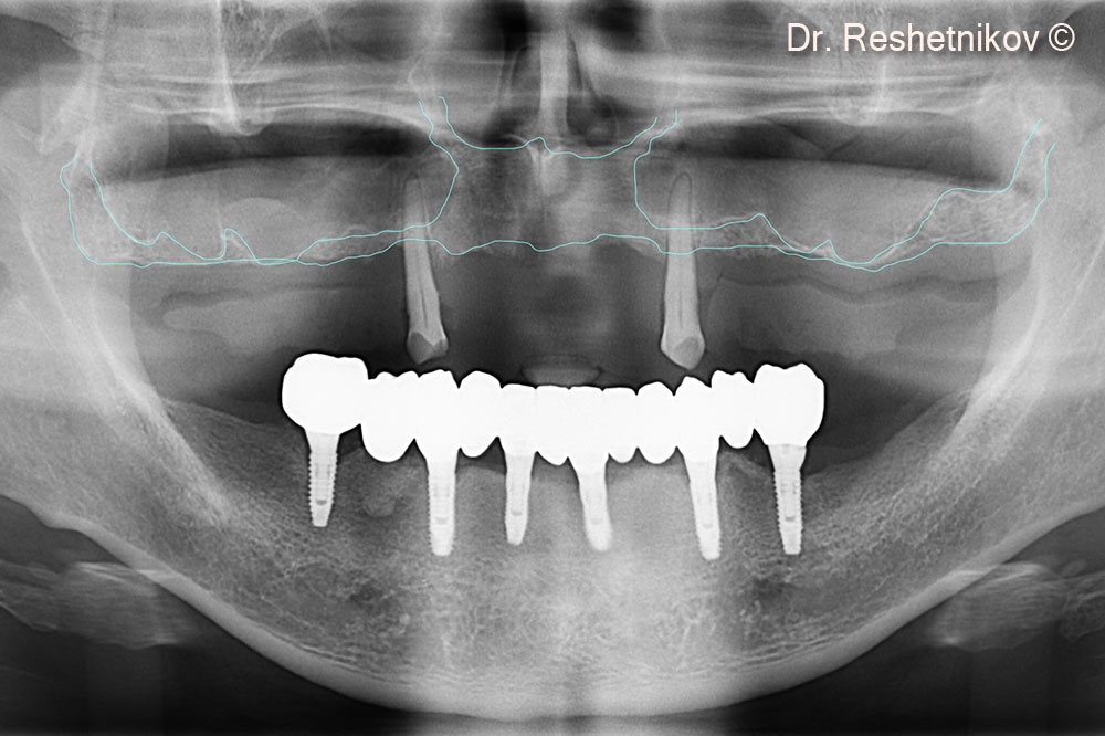
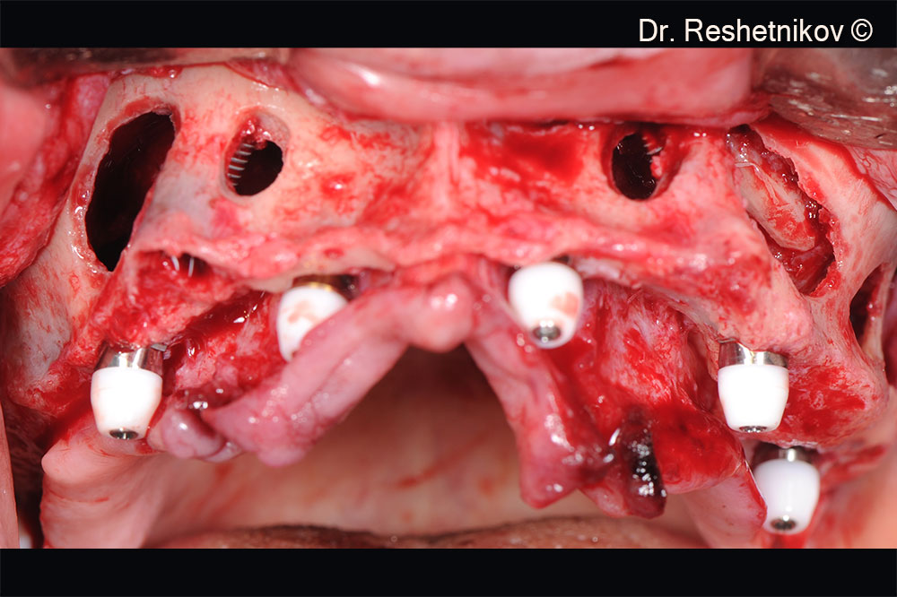
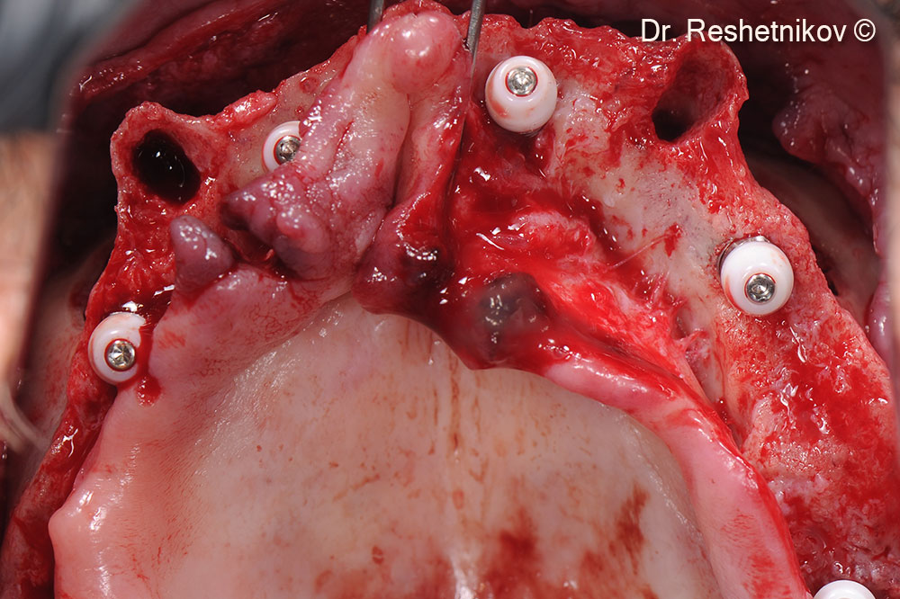
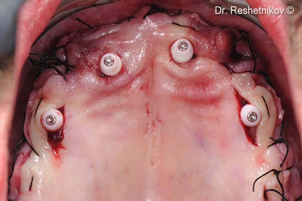
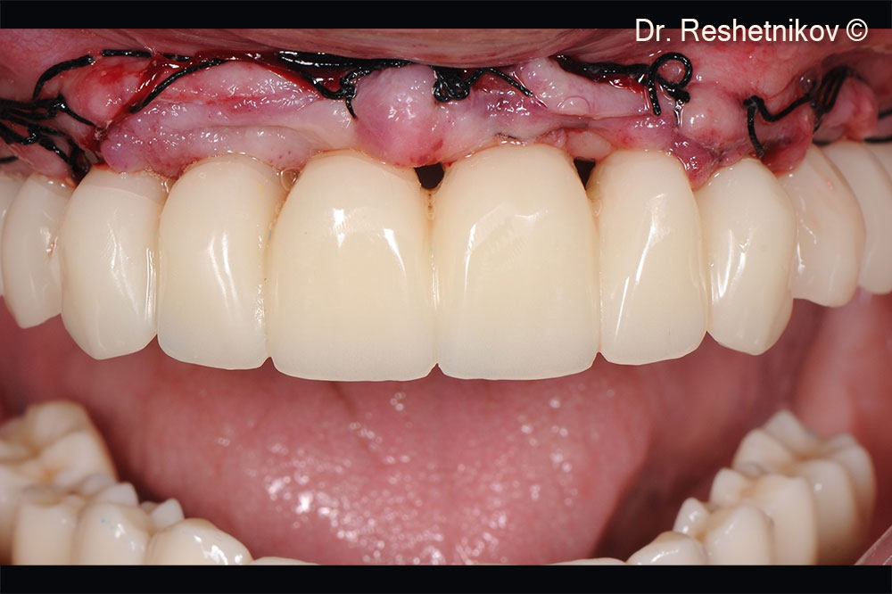
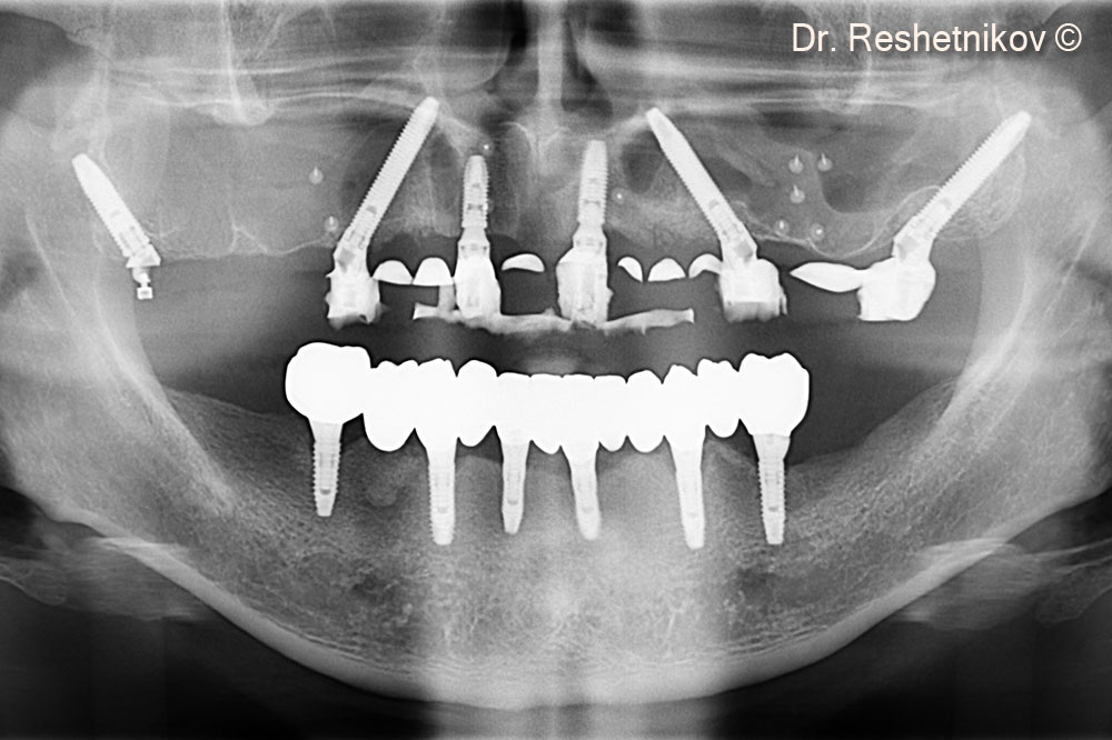
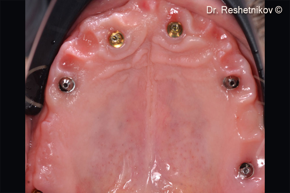
A male patient (58 years old) lacks teeth in the upper jaw.
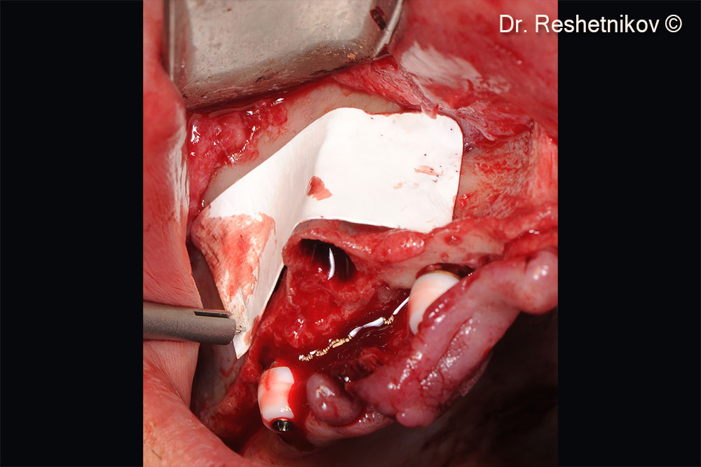
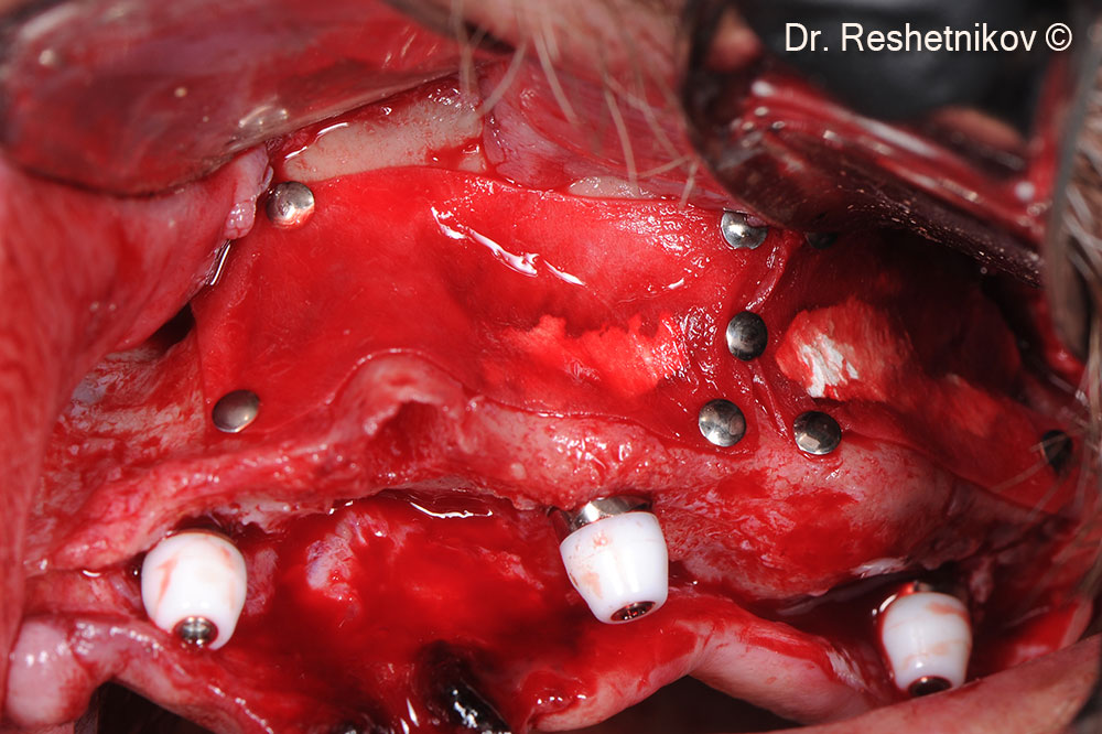
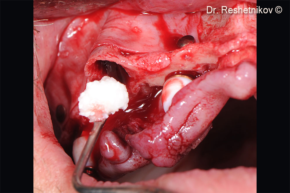
- mp3®
- Evolution

Lack of teeth in the upper jaw and aesthetic dissatisfaction with the removable denture

Alveolar ridges of the upper jaw atrophied unevenly

Alveolar ridges of the upper jaw atrophied unevenly

Panoramic radiograph shows pronounced pneumatization of the maxillary sinuses and atrophy of the alveolar ridge of the maxilla. Their edges highlighted in blue

After infiltration anesthesia, teeth #13 and #23 removed, vestibular incision made and the mucoperiosteal flap skeletonized. Formed access to the anterior and lateral walls of both maxillary sinuses with mobilization of the Schneiderian membrane

Inclined implants installed transsinusally using the “All-on-4” protocol

Void between the anterior wall of maxillary sinus and the Schneiderian membrane filled with mp3®. Bilateral approaches to the maxillary sinuses were closed with the collagen membrane Evolution

Void between the anterior wall of maxillary sinus and the Schneiderian membrane filled with mp3®. Bilateral approaches to the maxillary sinuses were closed with the collagen membrane Evolution

To prevent retraction of the socket walls it was filled with mp3®

Four holes made in the palatal flap to uncover the abutments and improve the quality of soft tissues around them. Suturing around installed abutments

Temporary prosthesis fixed to achieve aesthetic and functional results and to form adequate occlusion on the day of surgery

Final panoramic radiograph after implantation in the upper jaw

Soft tissues 6 months post-op

Installation of a permanent prosthesis using the FP2 protocol

Installation of a permanent prosthesis using the FP2 protocol

Panoramic radiograph 6 months after implantation with a permanent prosthesis installed on the upper jaw. The new edges of maxillary sinuses and alveolar ridge of the upper jaw highlighted in blue