The Modified Papilla Preservation Technique
Dr. Gerd Körner
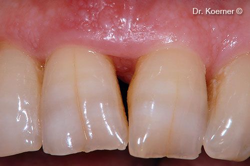
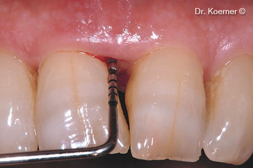
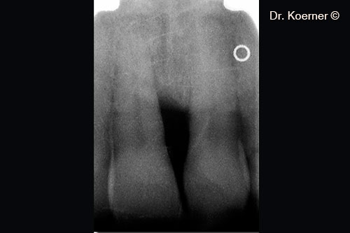
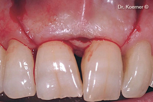
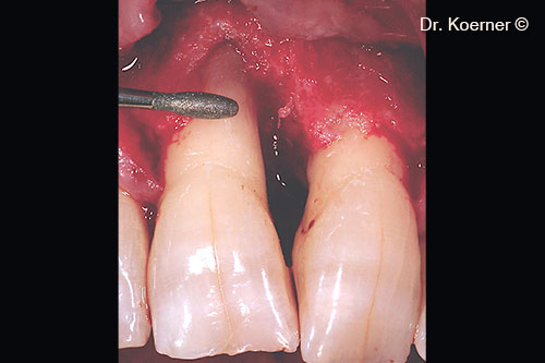
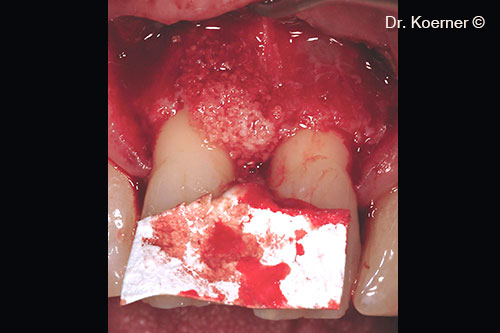
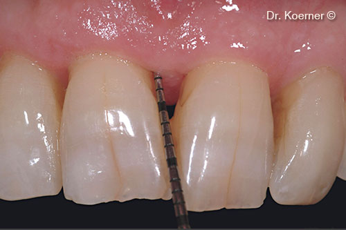
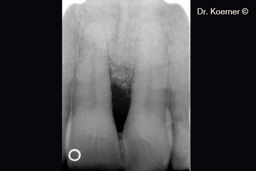
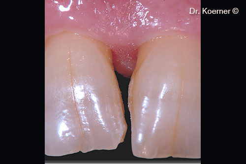
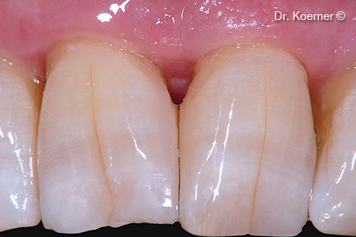
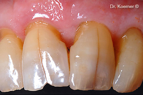
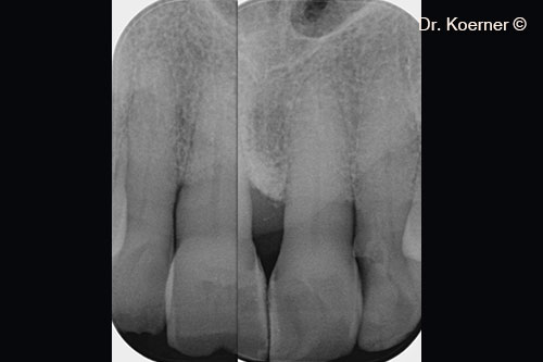
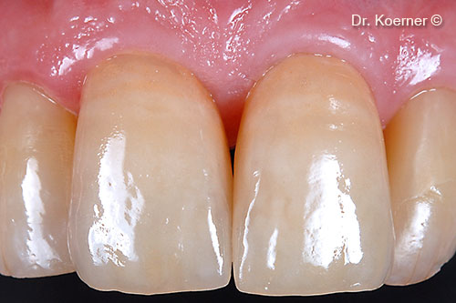
Initial situation
A male patient (61 years old) shows a periodontal defect in the esthetic area
OsteoBiol by Tecnoss
Attention please! The OsteoBiol® website contains information on Medical Devices, which may be dangerous for the patient health and safety if not used exclusively by medical professionals.