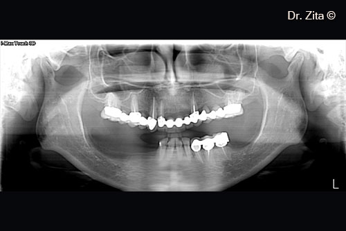
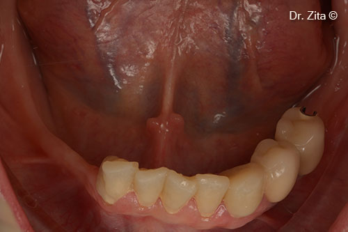
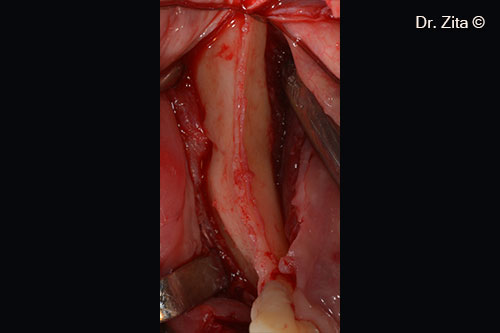
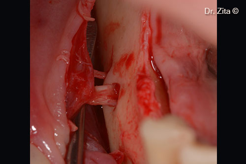
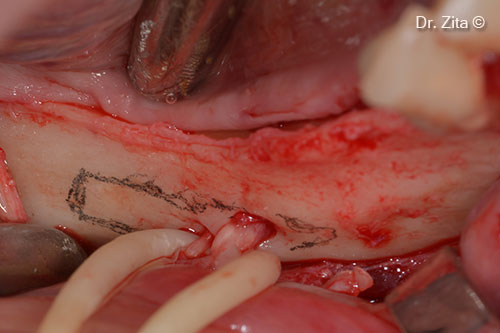
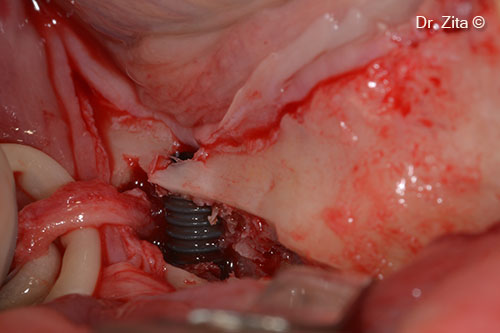
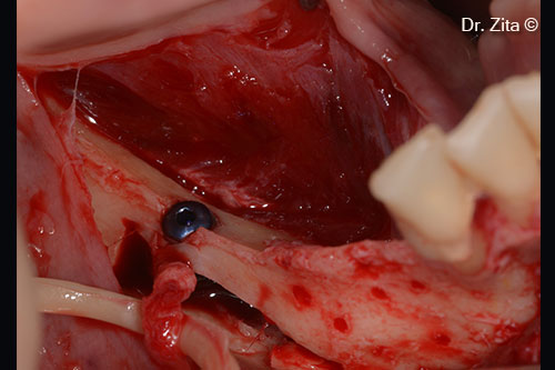
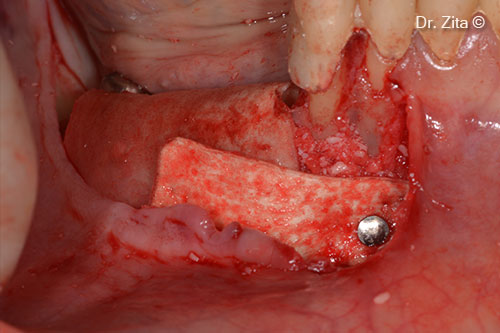
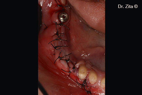
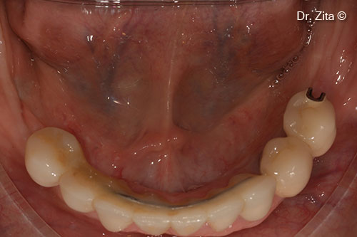
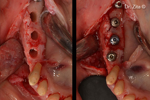
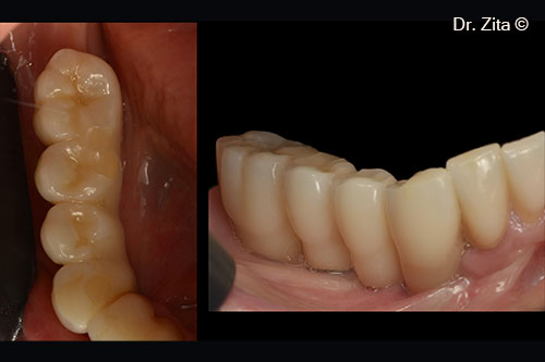
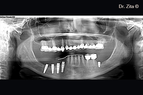
A female patient (43 years old) shows the mental foramen near the bone ridge
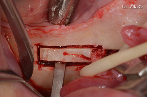
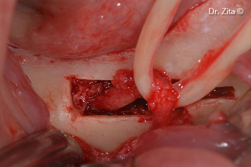
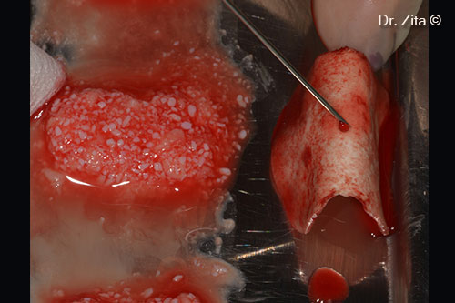
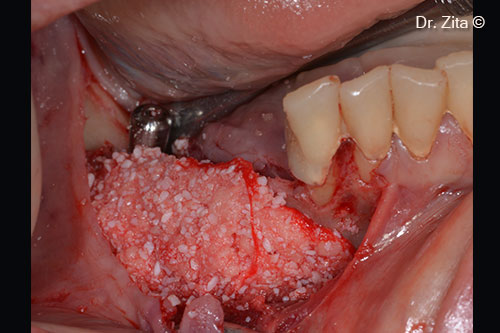
- mp3®
- Putty
- Lamina®
1. The initial panoramic x-ray shows the mental foramen near the bone ridge
2. Initial clinical photographic intra-oral situation
3. The bone ridge is exposed after a full thickness flap is rised. Tissues are incised with a 15c scalpel blade
4. Mental (main and accessory) nerves are exposed and individualized
5. Mental nerves are retracted with yellow sterile rubber. A window for exposing the inferior alveolar and incisive nerves is designed , with grey sterile pencil
6. After cutting the window to expose the IAN with piezotome, the cortical wall is exposed with the help of a chisel
7. The IAN nerve is removed from his canal after cutting the incisive part of the nerve 3 mm in front of the mental nerve (IAN transposition)
8. An implant is placed in front of new position of transposed IAN
9. The bone that needs to be regenerated horizontally, mesially to transposed IAN is decorticated.
10. mp3® is mixed with autologous bone (plate from the window that is milled with bone mill) and I-PRF and Lamina® Curved with I-PRF
11. Sticky bone (mp3®, autologous bone and I-PRF) in place
12. Lamina® Soft is placed after placing some Putty between the sticky bone and the Lamina®. The Lamina® is also stabilised by a metallic pin and with sutures
13. Soft tissues are sutured with non resorbable sutures supramid 4(0)
14. Result after 6 months of healing of the horizontal ridge augmentation
15. Three implants are placed on the regenerated ridge
16. Panoramic X-ray with all implants placed
17. Final rehabilitation with metalo - ceramic crowns. Case with seven years of follow-up