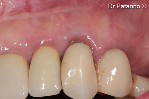
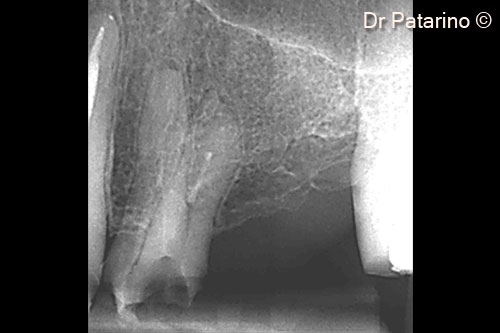
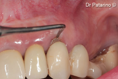
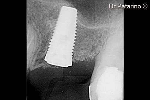
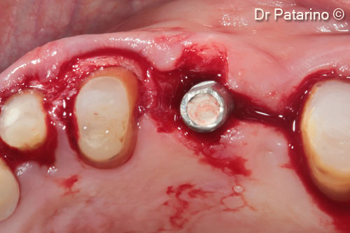
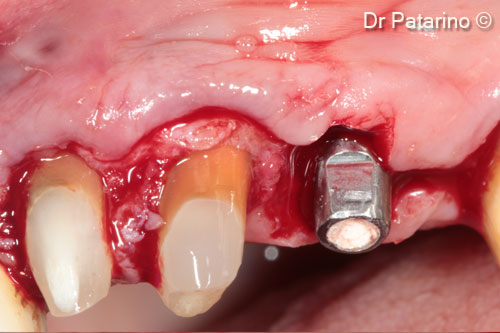
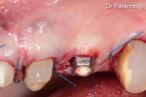
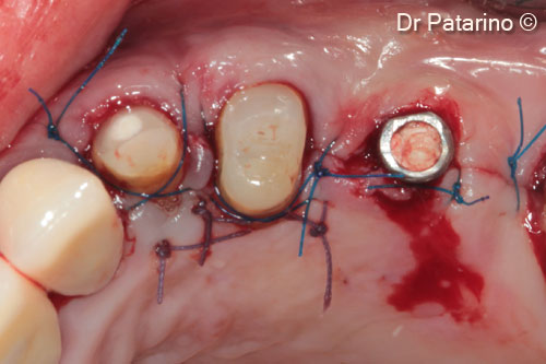
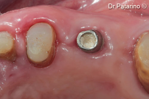
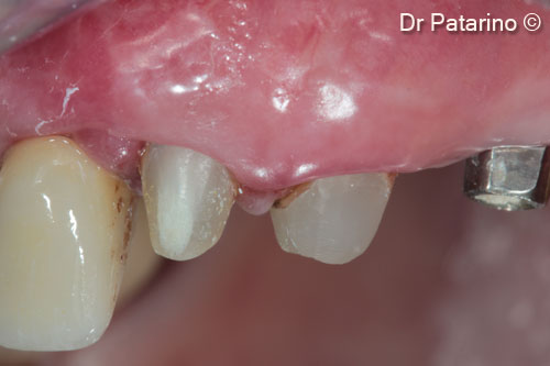
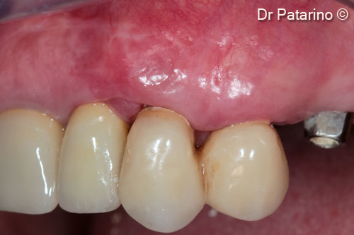
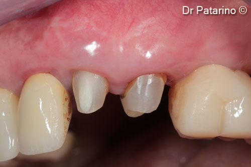
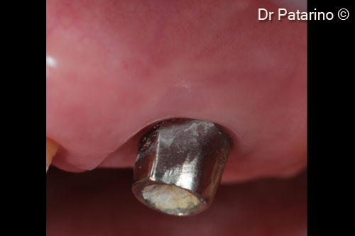
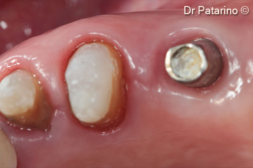
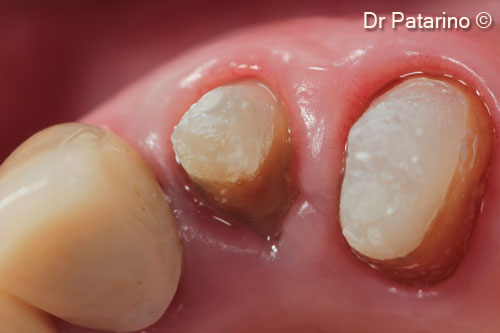
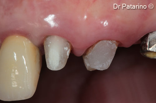
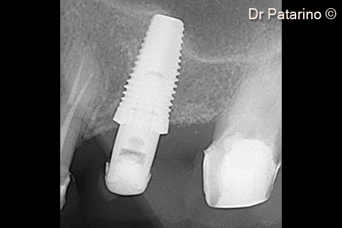
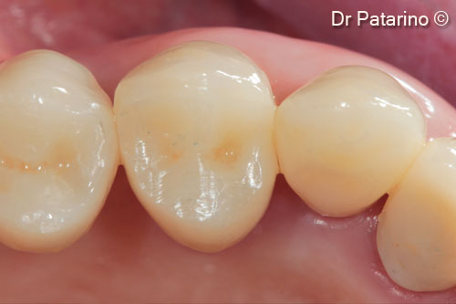
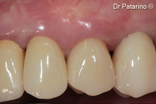
Initial situation
A female patient (56 years old) shows a hopeless tooth (upper jaw)
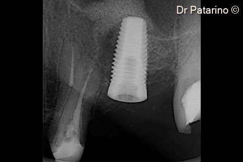
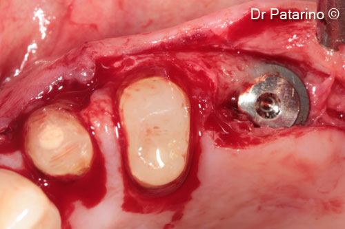
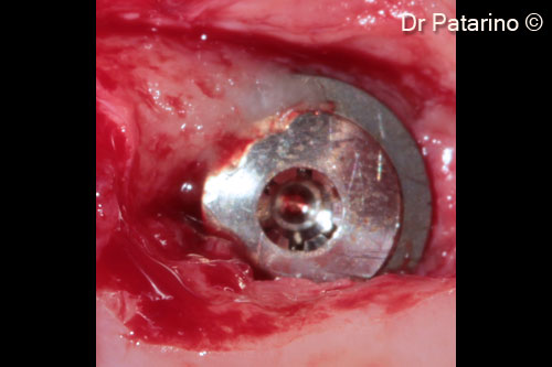
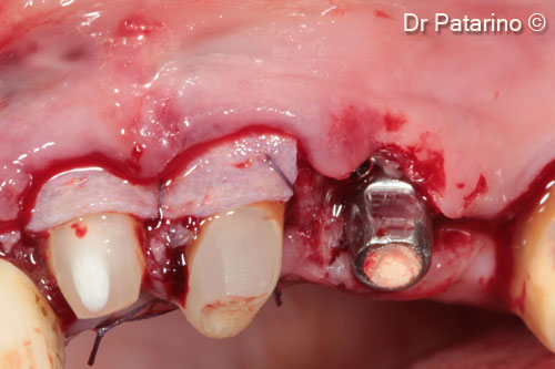
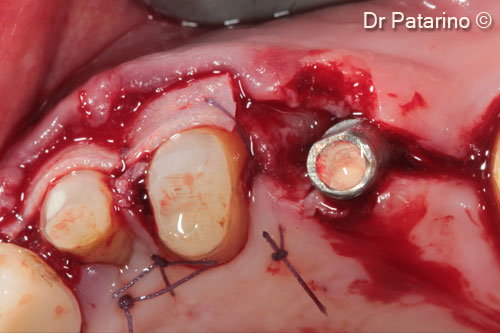
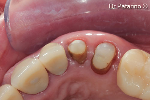
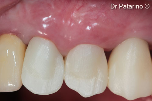
Used biomaterials
- Derma
- Evolution
Attention please! The OsteoBiol® website contains information on Medical Devices, which may be dangerous for the patient health and safety if not used exclusively by medical professionals.