A female patient (33 years old) requires ridge augmentation in the upper jaw.
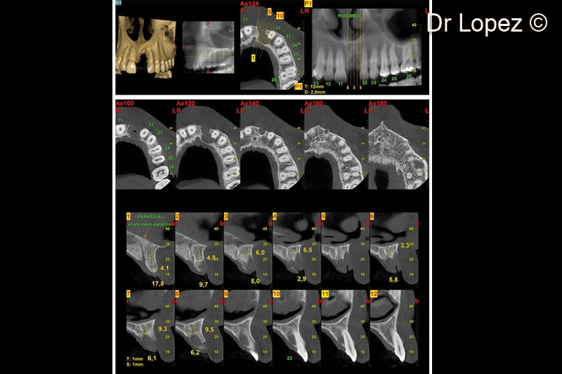
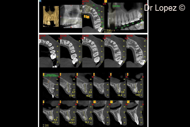
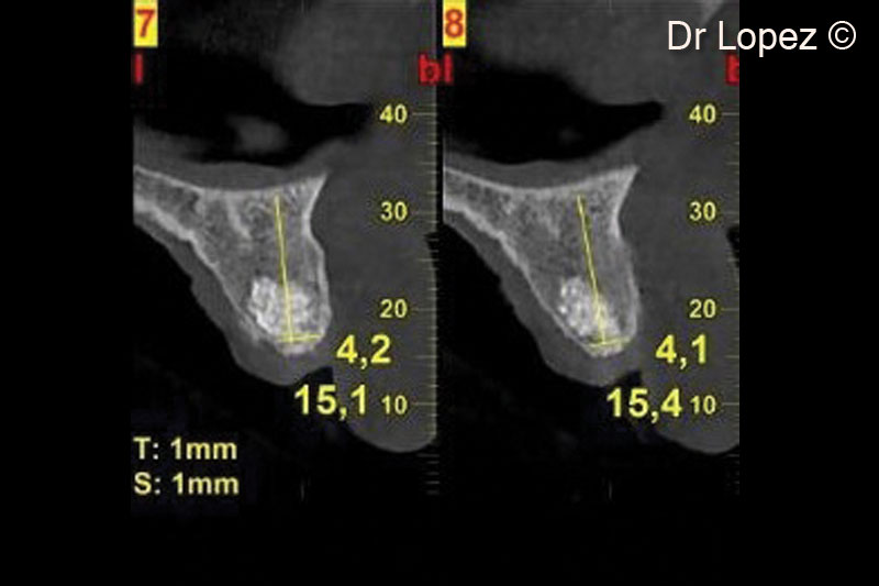
1. Pre-operative cone beam scan
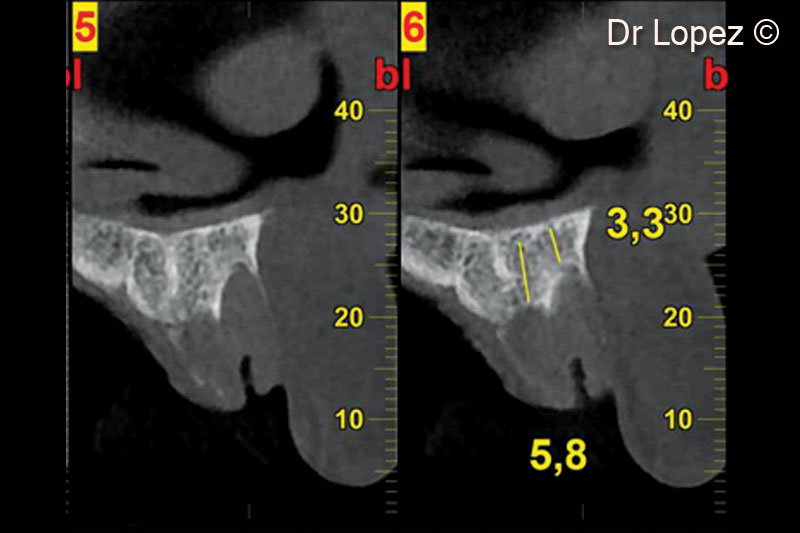
2. Pre-operative cone beam scan
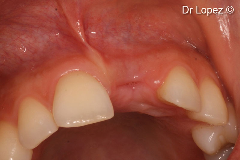
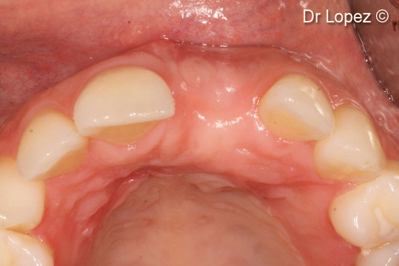
3. Post-traumatic bone defect
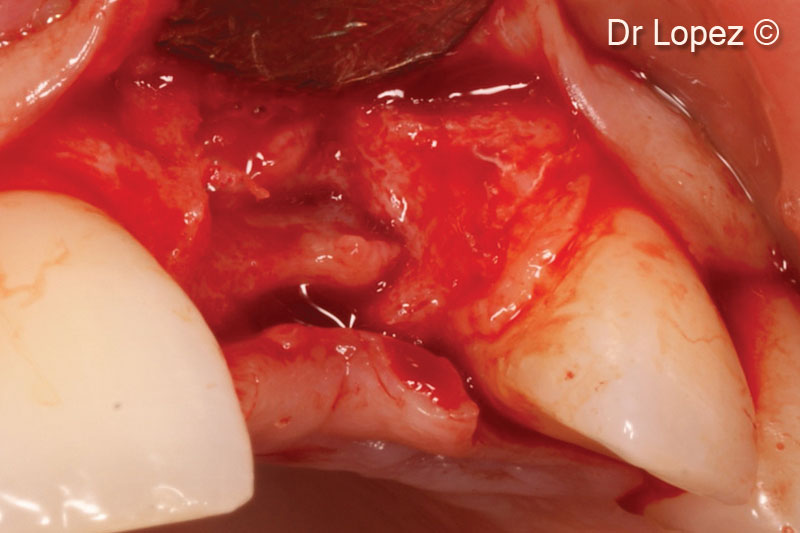
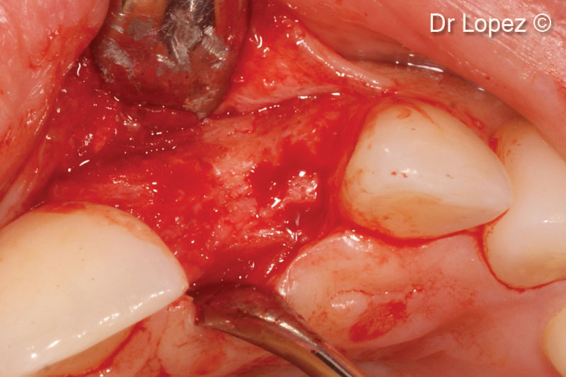
4. Flap opening
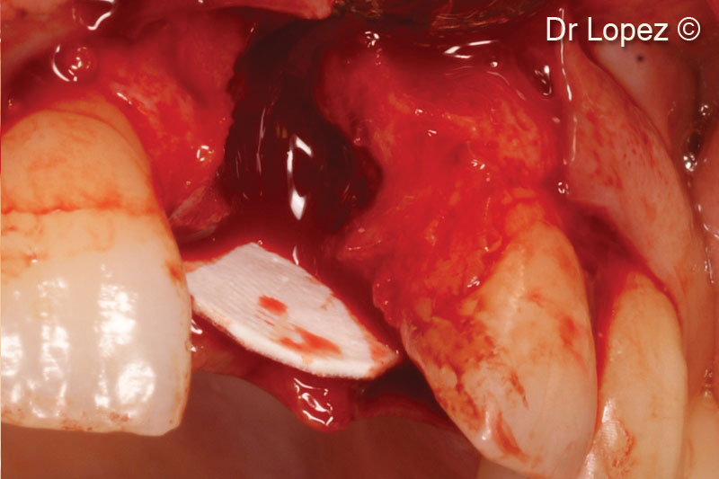
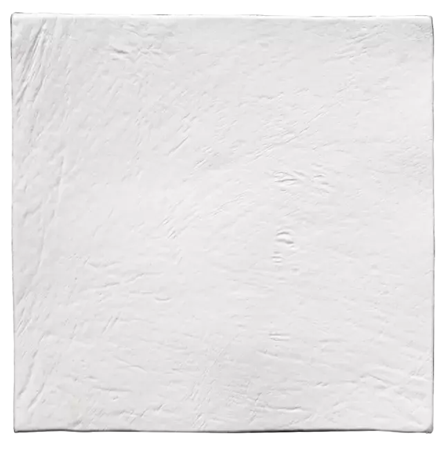
5. 1-mm thick OsteoBiol® Lamina® Soft positioned palatally
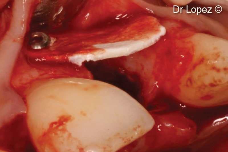
6. Fixation of the OsteoBiol® Lamina® Soft on the vestibular bone with titanium pins
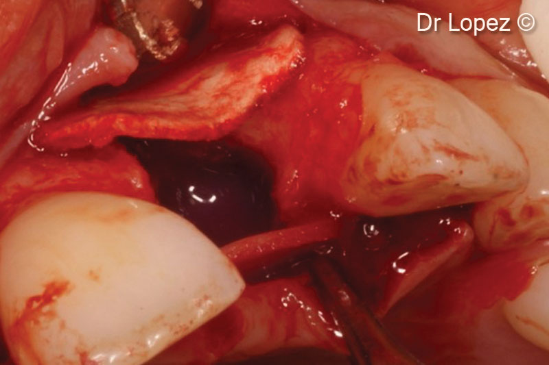
7. View of the defect grafted with OsteoBiol® Lamina® Soft
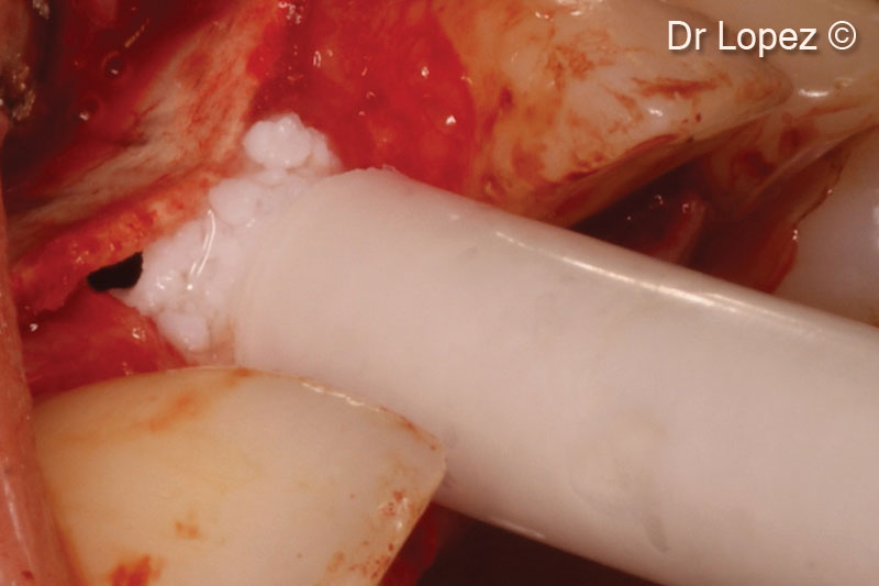
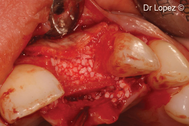
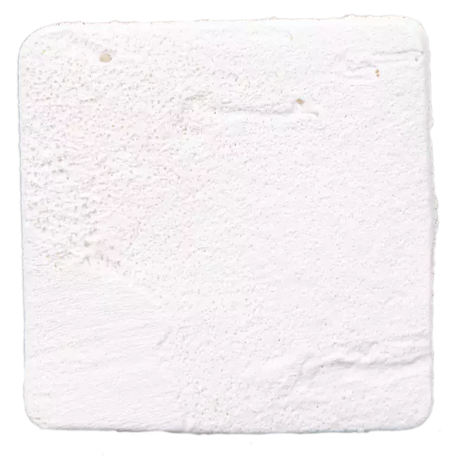
8. Grafting of OsteoBiol® mp3®
9. Defect completely filled
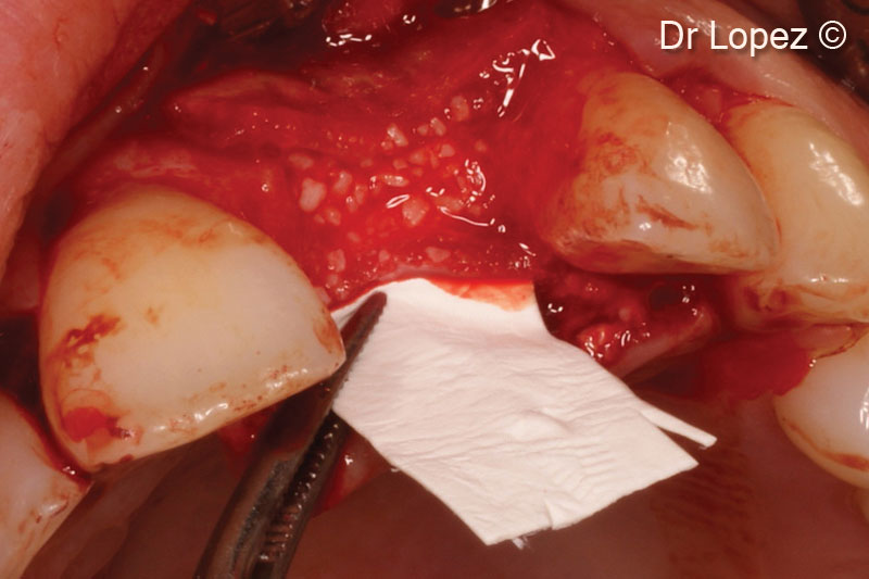
10. Covering of the grafted area with OsteoBiol® Evolution membrane
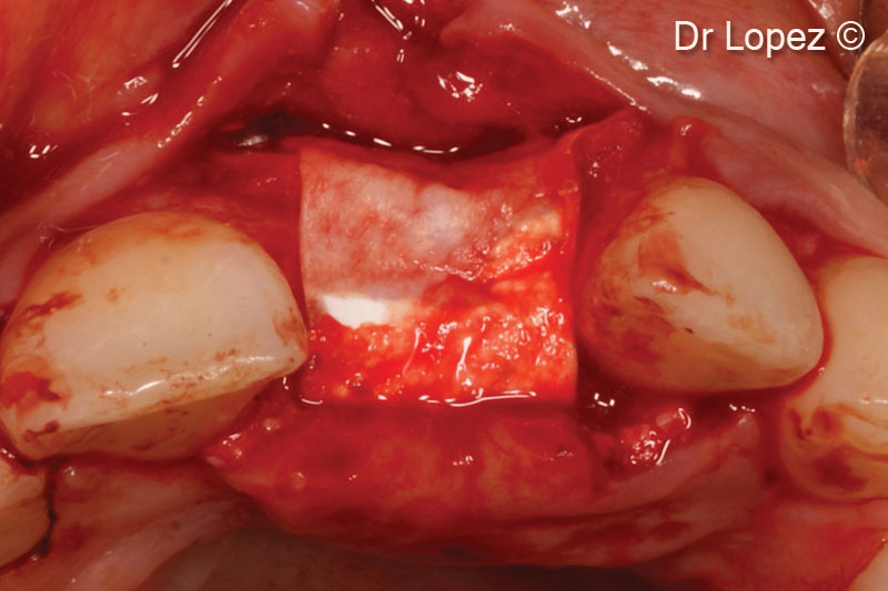
11. Final view of the grafted area
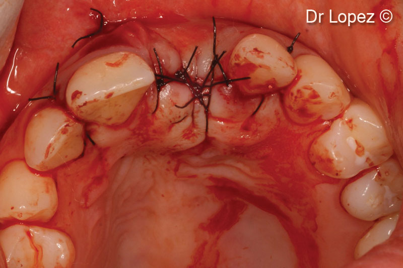
12. Flap closure
13. Post-operative cone beam scan at implant placement (8 months after surgery)
14. Post-operative cone beam scan at implant placement (8 months after surgery)
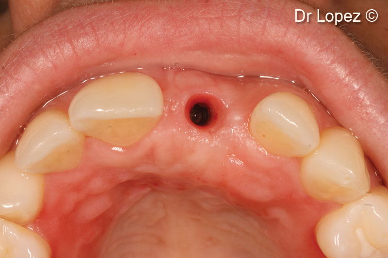
15. Pre-operative view of the augmented area
16. Intraoperative view
17. Positioning of a 3.5x10-mm implant
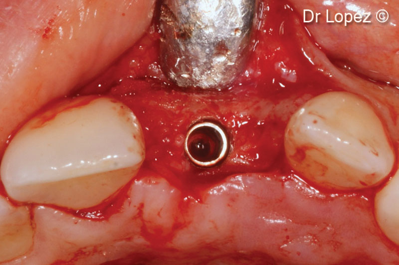
18. Tissue conditioning around implants
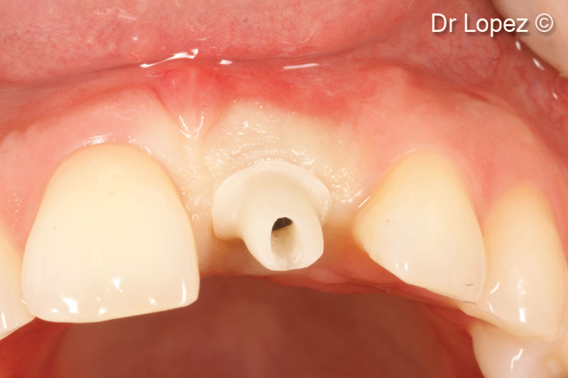
19. Ti-base abutment
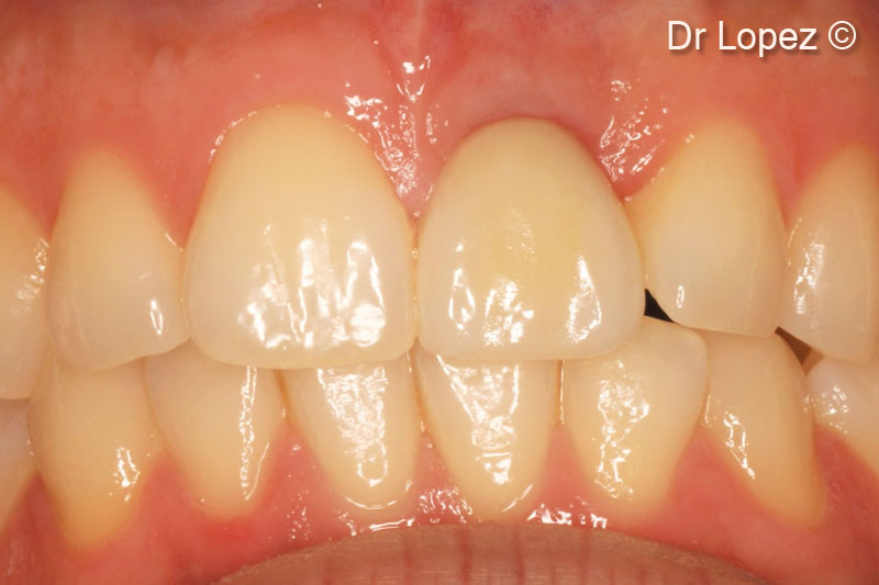
20. Control at 6 months
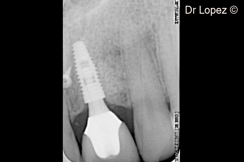
21. X-ray control at 3 years
Attention please! The OsteoBiol® website contains information on Medical Devices, which may be dangerous for the patient health and safety if not used exclusively by medical professionals.