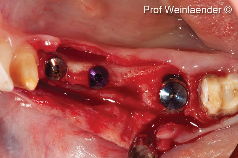
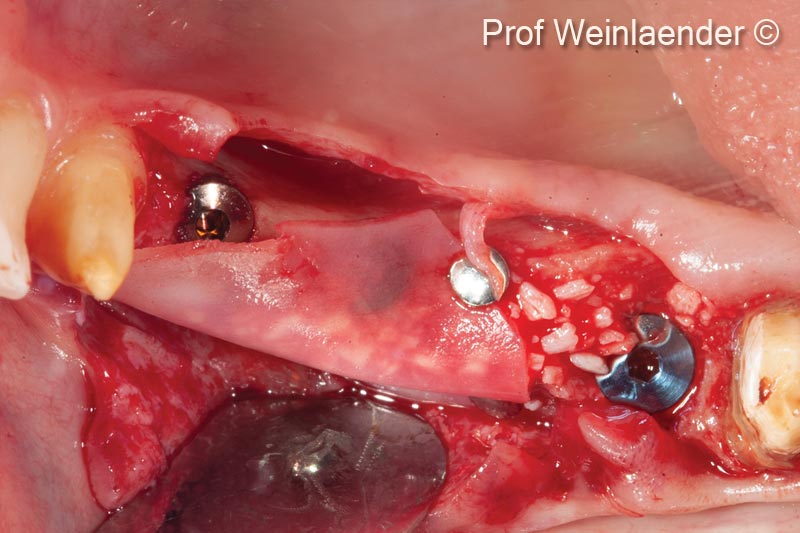
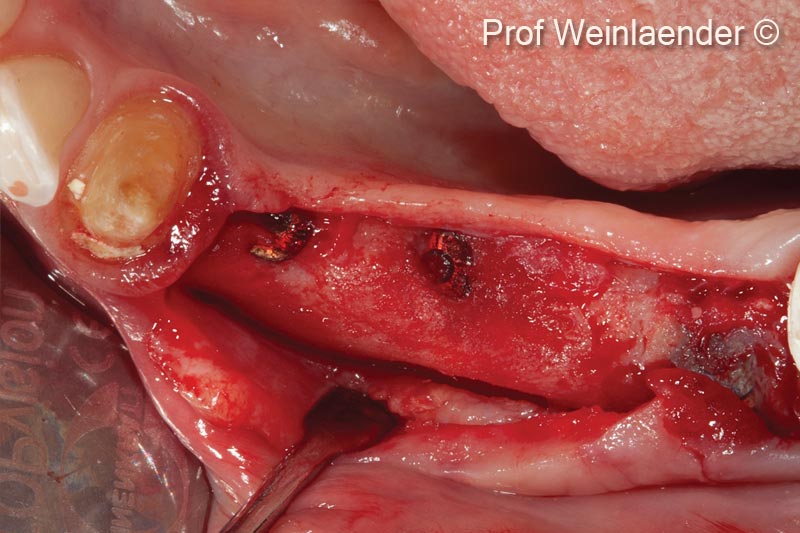
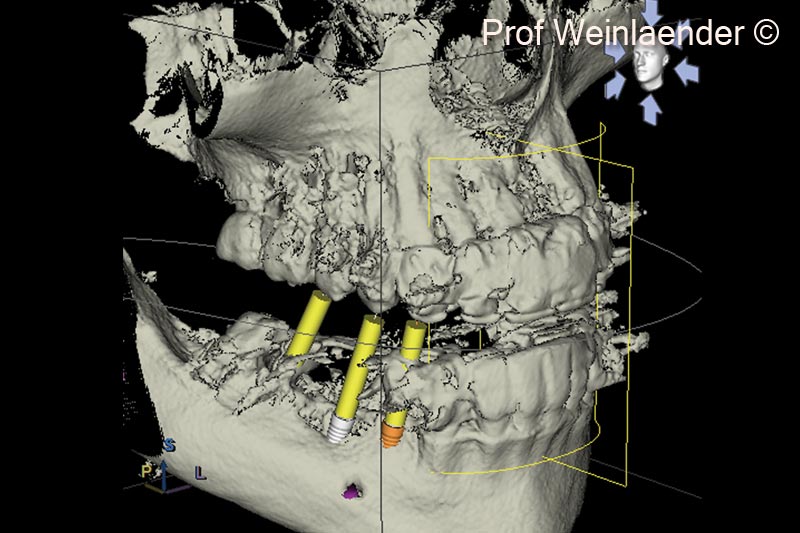
GBR at the same time of implant placement

Prof. Michael Weinländer
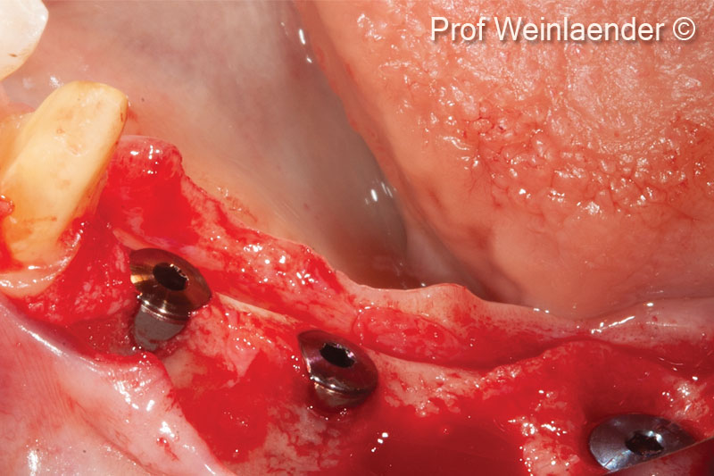
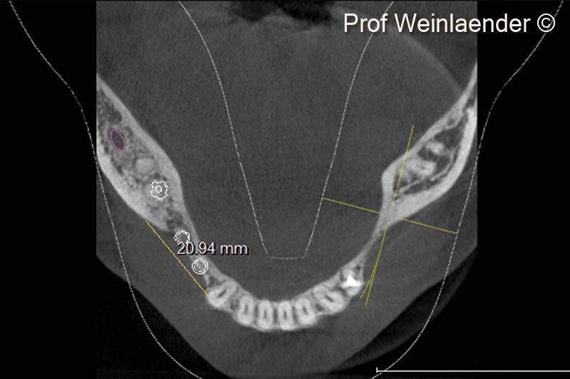
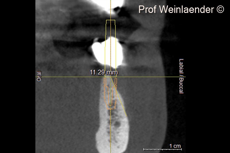
Initial situation

A female patient (43 years old) shows three implants positioned in a narrow ridge

Used biomaterials
  • mp3®
  • Lamina®
1. Positioning of 3 implants in position #44 #45 #46. #44 and #45 situated in a narrow ridge, #46 immediate implantation - Lateral view

1. Positioning of 3 implants in position #44 #45 #46. #44 and #45 situated in a narrow ridge, #46 immediate implantation - Lateral view

2. Positioning of 3 implants in position #44 #45 #46. #44 and #45 situated in a narrow ridge, #46 immediate implantation - Occlusal view

2. Positioning of 3 implants in position #44 #45 #46. #44 and #45 situated in a narrow ridge, #46 immediate implantation - Occlusal view

3. Placement of OsteoBiol® Lamina® with the creation of a stable lateral void through pin fixation at the height of tooth 43, the depth of the vestibulum, the occlusal part of the alveolar ridge and the filling of the created void with the use of OsteoBiol® mp3® (xenogenic pre-hydrated collagenated cortico-cancellous bone particles)

3. Placement of OsteoBiol® Lamina® with the creation of a stable lateral void through pin fixation at the height of tooth 43, the depth of the vestibulum, the occlusal part of the alveolar ridge and the filling of the created void with the use of OsteoBiol® mp3® (xenogenic pre-hydrated collagenated cortico-cancellous bone particles)

4. Situation 8 months post-operative. Transformation of the GBR complex into a clinically successful augmentation not only covering the exposed coronal parts of implants but also extending the lateral aspect of the alveolar ridge for about 3-4 mm additionally

4. Situation 8 months post-operative. Transformation of the GBR complex into a clinically successful augmentation not only covering the exposed coronal parts of implants but also extending the lateral aspect of the alveolar ridge for about 3-4 mm additionally

5. Occlusal DVT view of implants in position #44 #45 #46 with the desired augmentative planning indicated by a straight line

5. Occlusal DVT view of implants in position #44 #45 #46 with the desired augmentative planning indicated by a straight line

6. Occlusal DVT view of implants in position #44 #45 #46 with the obtained augmentation indicated by a straight line

6. Occlusal DVT view of implants in position #44 #45 #46 with the obtained augmentation indicated by a straight line

7.  Radial DVT view of the implant in position 44 indicating the amount of alveolar ridge resorption at the planned implant position

7. Radial DVT view of the implant in position 44 indicating the amount of alveolar ridge resorption at the planned implant position

8. Radial DVT view of the implant in position #45 indicating the amount of alveolar ridge resorption at the planned implant position

8. Radial DVT view of the implant in position #45 indicating the amount of alveolar ridge resorption at the planned implant position

9. 3D Surface rendering DVT. Demonstration of narrow ridge with exposed implants in positions #44 and #45

9. 3D Surface rendering DVT. Demonstration of narrow ridge with exposed implants in positions #44 and #45

Used biomaterials
mp3
mp3®
lamina
Lamina®
Are you a medical professional?
No

Attention please! The OsteoBiol® website contains information on Medical Devices, which may be dangerous for the patient health and safety if not used exclusively by medical professionals.