A female patient (45 years old) shows a deficient bone ridge
- Evolution
- Gen-Os®
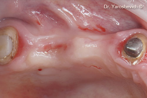
Initial intraoral view. Site with a ridge contour deficit
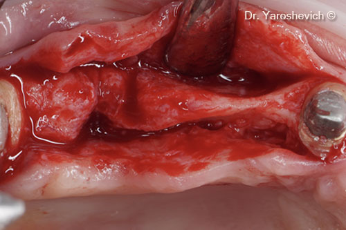
In the posterior maxilla, a full-thickness, slightly buccal incision was in the keratinized gingiva
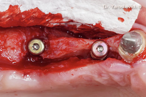
Two implants successfully placed at sites #14 and #16, with good primary stability. Instead of a cover screw, a 3-mm healing abutment was used
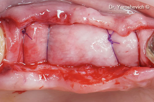
Evolution collagen membrane fixed with a resorbable suture material (no pins needed to stabilize it) and adapted to extend 2 mm beyond the defect margins
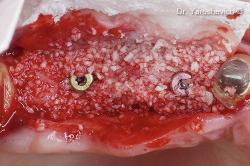
Collagenated cortico-cancellous bone mix Gen-Os® placed buccally and lingually on the defect site
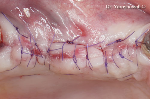
Soft tissues sutured with double simple 6/0 sutures
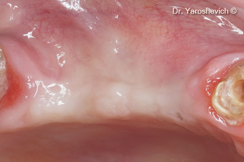
Intraoral view after 6 months
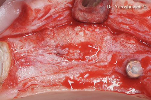
Full-thickness flap elevated (during the second stage). Increase in bone width observed 6 months after augmentation
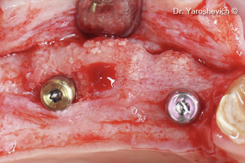
3-mm healing abutments replaced with 5-mm healing abutments
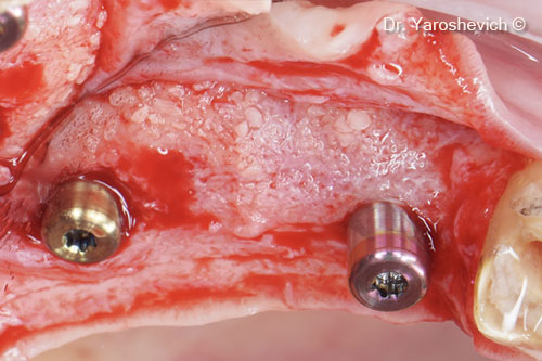
Homogenous bone around the healing abutment with 8-mm of bone width observed
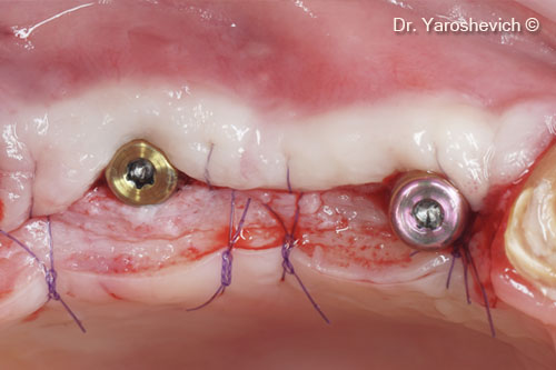
Soft tissues sutured with figure 8 sutures
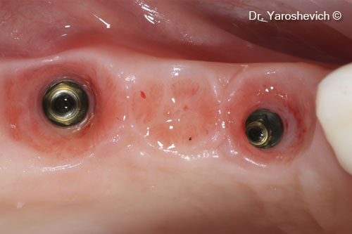
Postoperative occlusal view
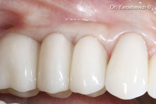
In 2 months, the patient underwent prosthetic treatment after a screw-retained zirconia crowns had been made
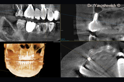
After 2 years, a new CBCT scan performed to evaluate the alveolar ridge around #14 implant horizontally
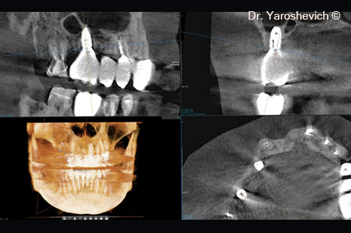
After analyzing the alveolar ridge horizontally, as observed 2 years post-op, the bone width around the #16 implant remained stable
