A female patient (50 years old) shows pneumatization of both maxillary sinuses
- mp3®
- Putty
- Evolution
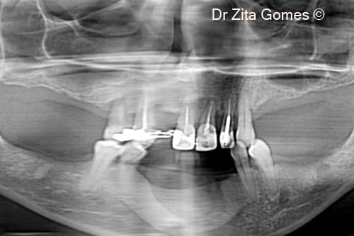
1. Initial panoramic x-ray, showing pneumatization of both maxillary sinuses
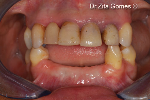
2. Initial intra-oral situation
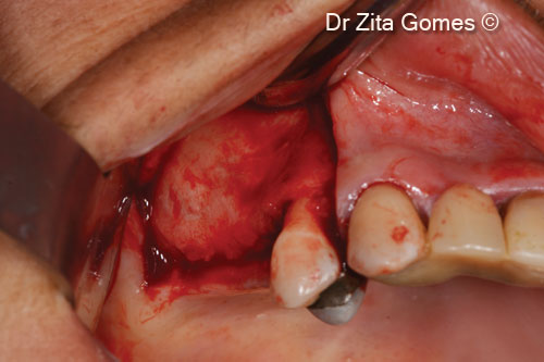
3. A full thickness mucoperiosteal flap, with mesial release incision, is raised to access the right maxillary sinus
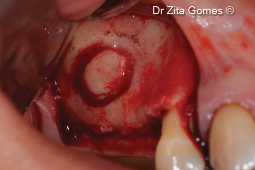
4. A lateral window is opened with a high speed bur
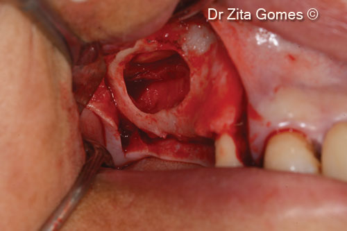
5. The Schneiderian membrane is raised to create the space for the graft
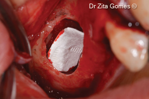
6. An Evolution membrane is used to protect the ceiling of the sinus
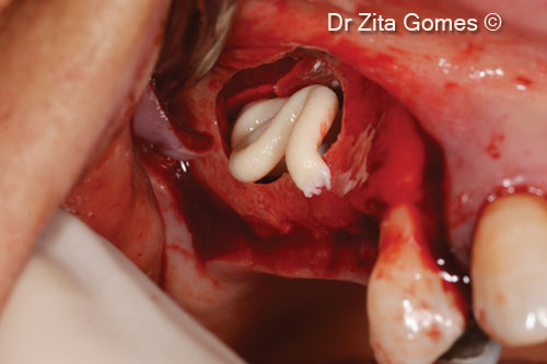
7. Beginning filling the lifted sinus with Putty
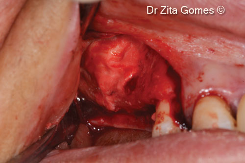
8. Finishing the filling of the sinus with Putty
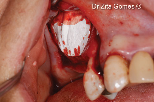
9. The access window is protected with an Evolution membrane
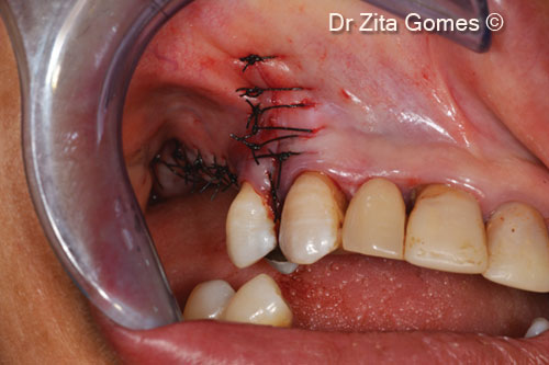
10. The flap is sutured with polyamide 4(0) suture with simple stitches
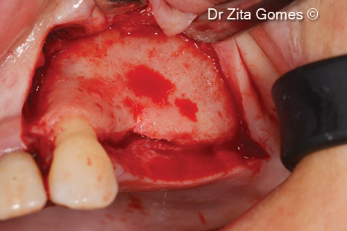
11. A full-thickness mucoperiosteal flap with mesial release incision is cretaed to access the left maxillary sinus
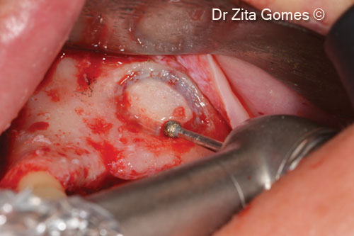
12. A lateral window is opened with a high speed bur
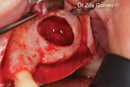
13. The Schneiderian membrane is raised to create the space for the graft
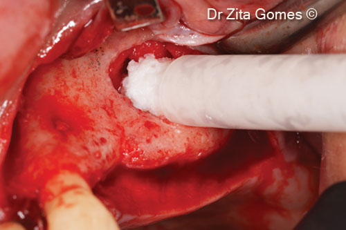
14. Beginning filling the lifted sinus with mp3®
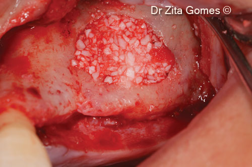
15. Finishing the filling of the sinus
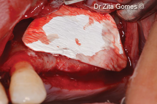
16. The access window is protected with an Evolution membrane

17. The flap is sutured with polyamide 4(0) suture with simple stitches

18. Four dental implants (2 in each sinus) are placed after eight months of healing. Rehabilitation with fixed ceramic bridges after three months

19. Final fixed oral rehabilitation with bridges over implants in the posterior regions and ceramic crows on the anterior teeth (occlusal view)

20. Patient smile after the rehabilitation

21. Panoramic final X-ray at five-year follow up


