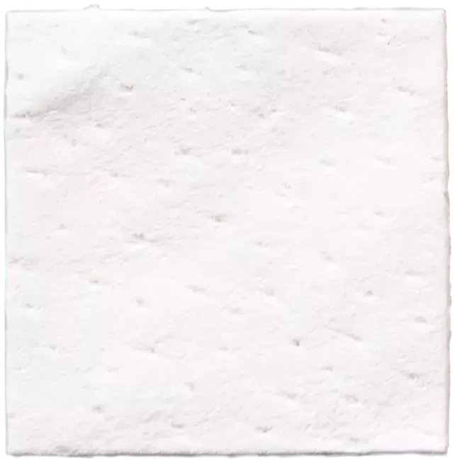
A male patient (64 years old) shows a gingival recession
- Derma
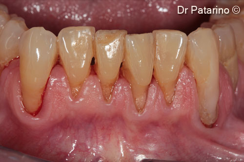
1. Lower jaw: gingival recessions in the anterior region
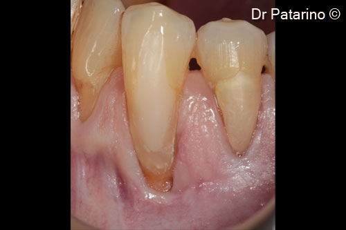
2. Dental element 3.3: 10-mm Miller class II recession from CEJ
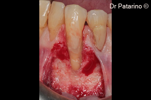
3. Partial thickness trapezoidal flap and papillar dissection
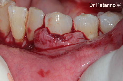
4. Coronal mobilization of the flap
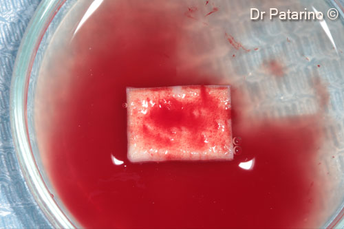
5. Derma membrane hydrated with saline and blood
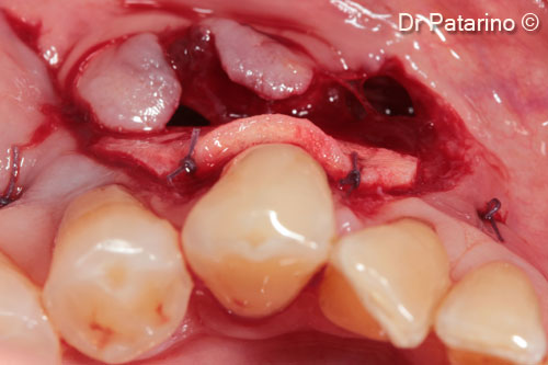
6. Occlusal view of the graft sutured at the receiving site
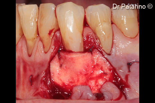
7. Derma graft, shake and apically sutured to CEJ and stabilized laterally with resorbable suture to the receiving site
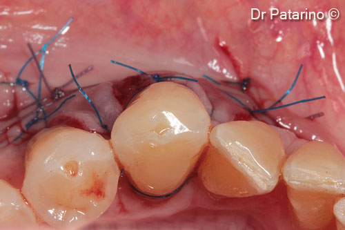
8. Coronally elevated flap to uncover the graft: occlusal view
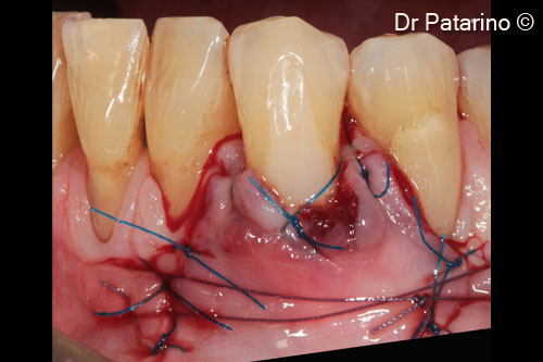
9. Sutures to enhance the adaptation of the coronal margin of the flap to the dental convexity, single sutures to stabilize the flap
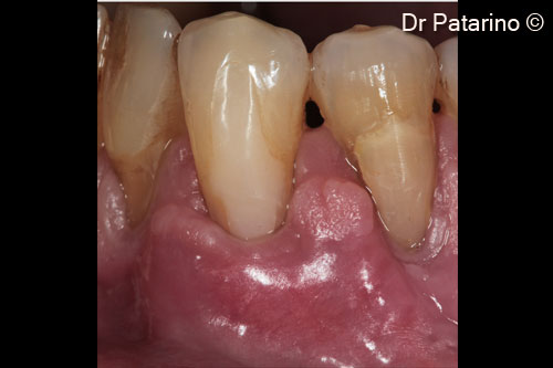
10. Healing at 20 days
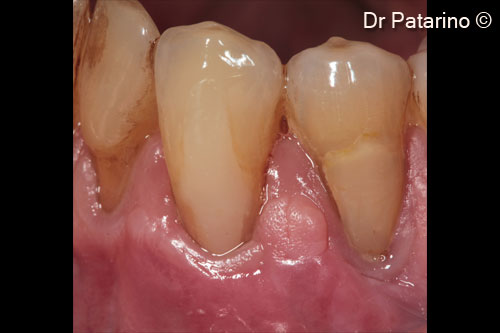
11. Healing at 2 months: good coverage of the roots and levelling of the muco-gingival line
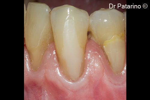
12. Healing at 9 months: good coverage of the roots and levelling of the muco-gingival line
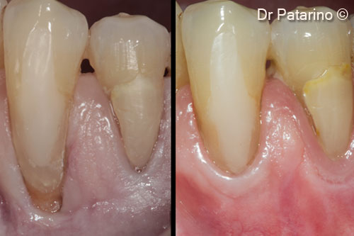
13. Comparison between the initial situation and the tissues at 9 months
