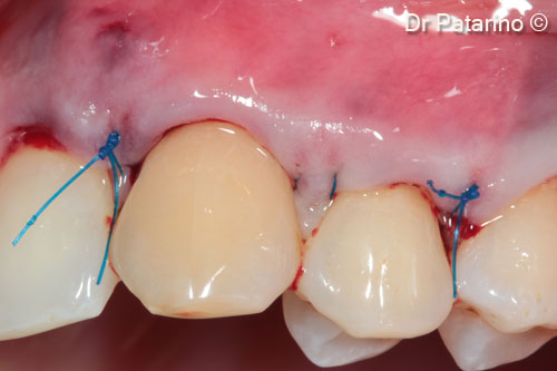
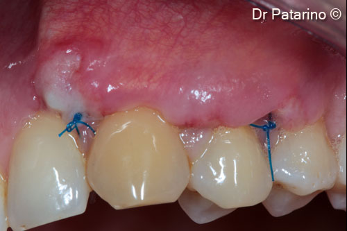
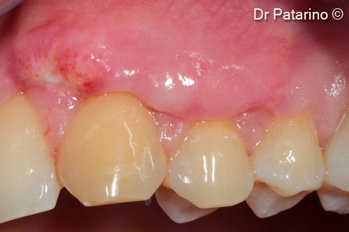
Bilaminar technique in multiple Miller Class I recessions
Dr. Domenico Patarino
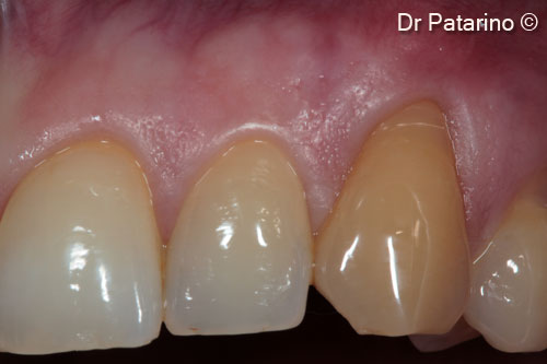
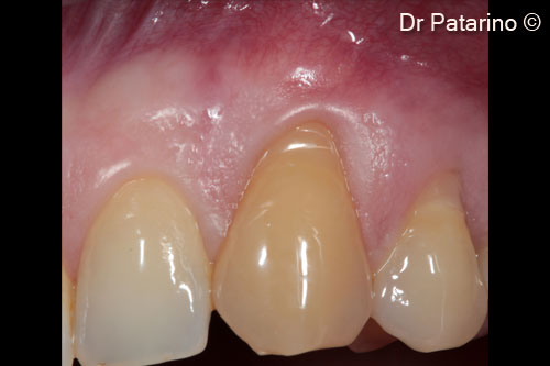
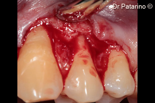
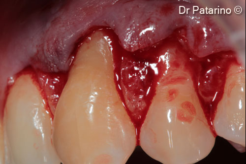
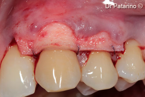
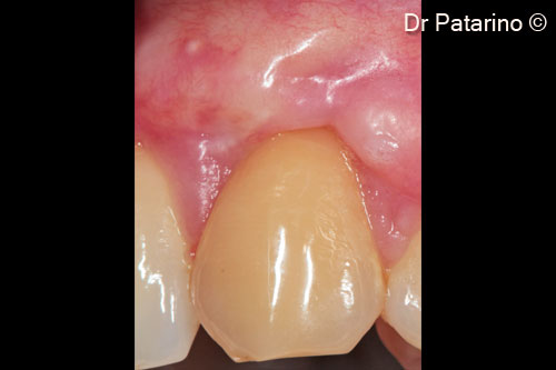
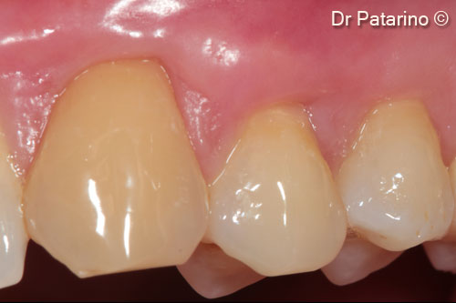
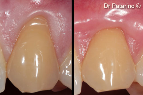
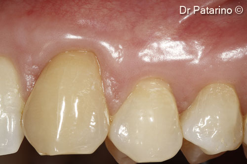
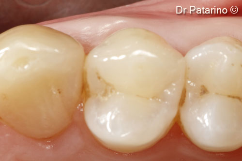
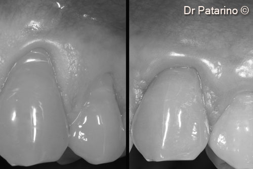
Initial situation
A female patient (35 years old) shows a Miller Class I gingival recession
OsteoBiol by Tecnoss
Attention please! The OsteoBiol® website contains information on Medical Devices, which may be dangerous for the patient health and safety if not used exclusively by medical professionals.