A female patient (46 years old) shows severe peri-implantitis around two implants
Used biomaterials
GTO®
1. The patient presented with severe peri-implantitis around 2 central incisors zirconia implants
2. The patient presented with severe peri-implantitis around 2 central incisors zirconia implants
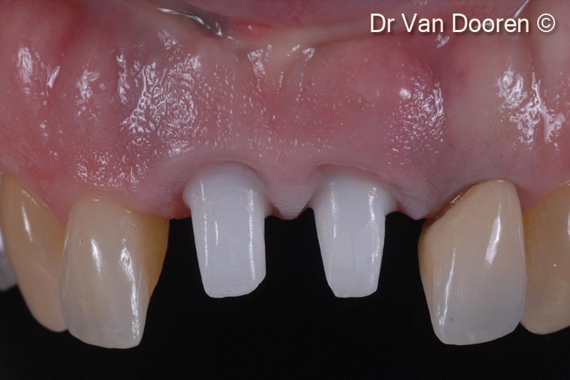
3. The old crowns were removed, and a provisional bridge was fabricated with a new design proposal. Pink composite was added to have a clear idea of the incisal edge position/toothform and artificial gingival replacement
4. The old crowns were removed, and a provisional bridge was fabricated with a new design proposal. Pink composite was added to have a clear idea of the incisal edge position/toothform and artificial gingival replacement
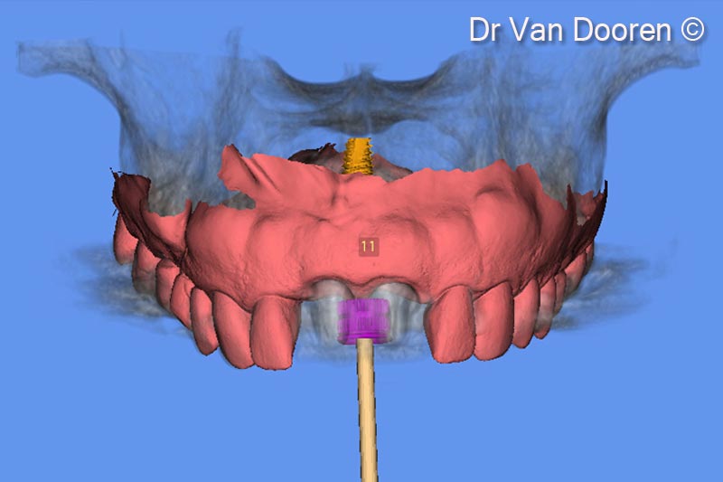
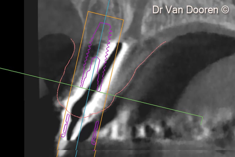
5. Superimposing the STL file and the Dicom files in the planning software (MSoft/MIS) allowed us to plan the exact position of the new implant
6. Superimposing the STL file and the Dicom files in the planning software (MSoft/MIS) allowed us to plan the exact position of the new implant
7. Superimposing the STL file and the Dicom files in the planning software (MSoft/MIS) allowed us to plan the exact position of the new implant
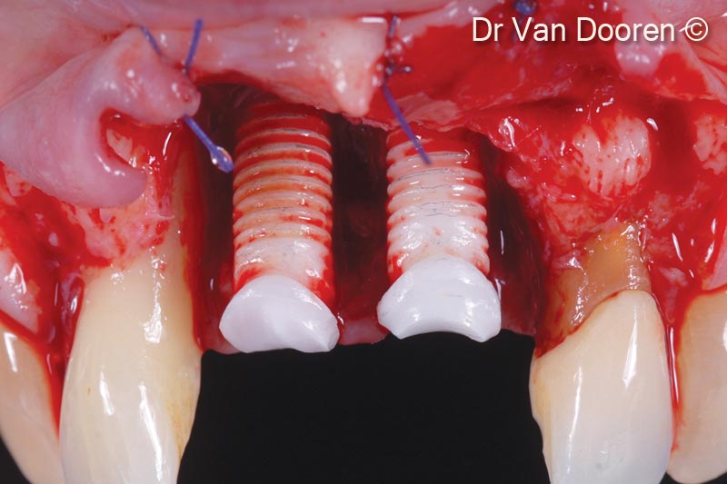
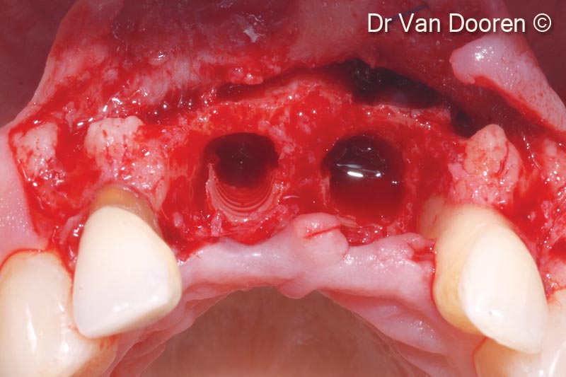
8. Considerable bone loss around the existing implants and mesially to the lateral incisors: the two implants were removed
9. Considerable bone loss around the existing implants and mesially to the lateral incisors: the two implants were removed
10. Occlusal view after implant removal: the ridge was surgically corrected and flattened after implant removal. The implant will be placed in a more palatal and “in between” position compared to the extraction sockets to get excellent primary stability for immediate loading
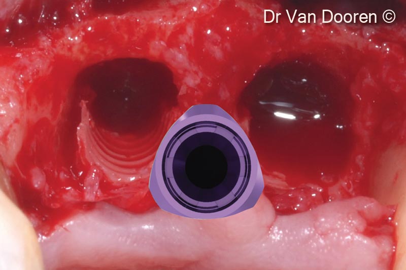
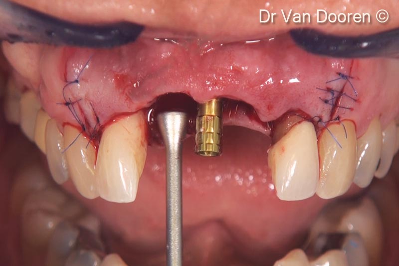
11. Placement of the implant (V3/ RP 3.9 mm - 13 mm) using the MGuide (MIS). The palatal position for obtaining a screw retained restoration is the key point in this specific case
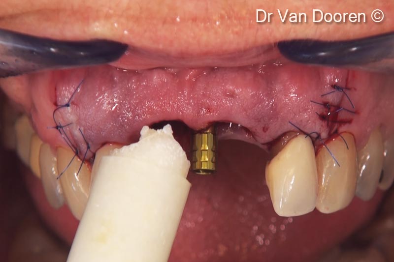
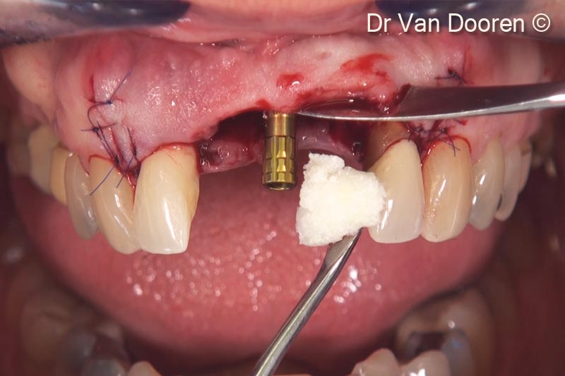
12. After placement of a provisional cylinder, OsteoBiol® GTO® was placed and packed, with the instruments and around the implant in order to support the soft tissue
13. After placement of a provisional cylinder, OsteoBiol® GTO® was placed and packed, with the instruments and around the implant in order to support the soft tissue
14. After placement of a provisional cylinder, OsteoBiol® GTO® was placed and packed, with the instruments and around the implant in order to support the soft tissue
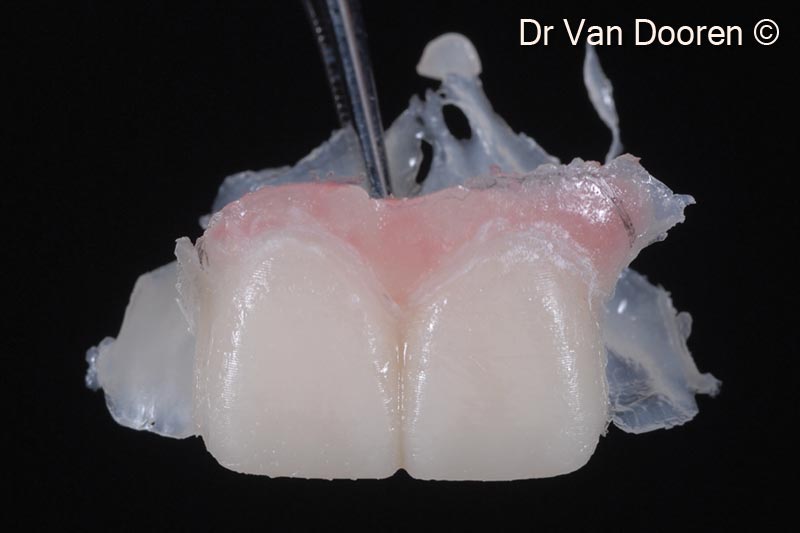
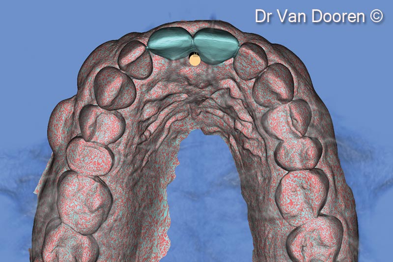
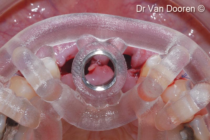
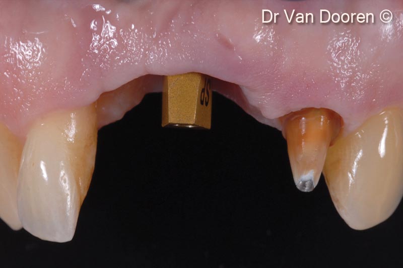
15. After making the final preparation of tooth #22, a digital impression was taken with a scanpost (MIS)
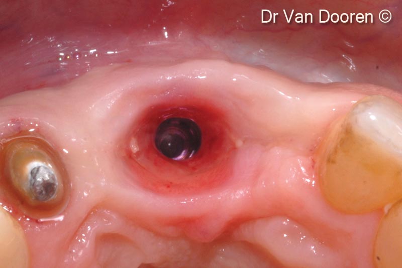
16. Occlusal view after 4 months. Please note the excellent soft tissue health and three-dimensional stability of the residual ridge after grafting
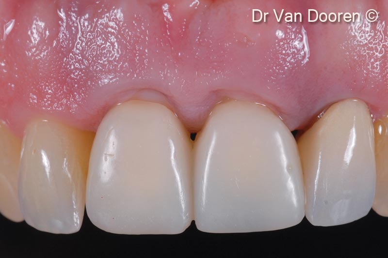
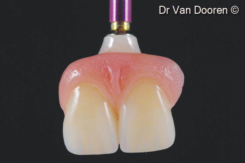
17. A 2-unit screw retained zirconia bridge (#11 and #21) and a single crown (tooth #22) were milled and layered
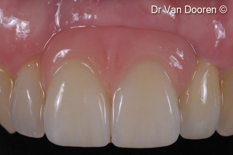
18. Final clinical result. Note the integration and gingival health
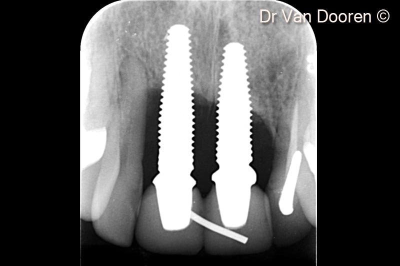
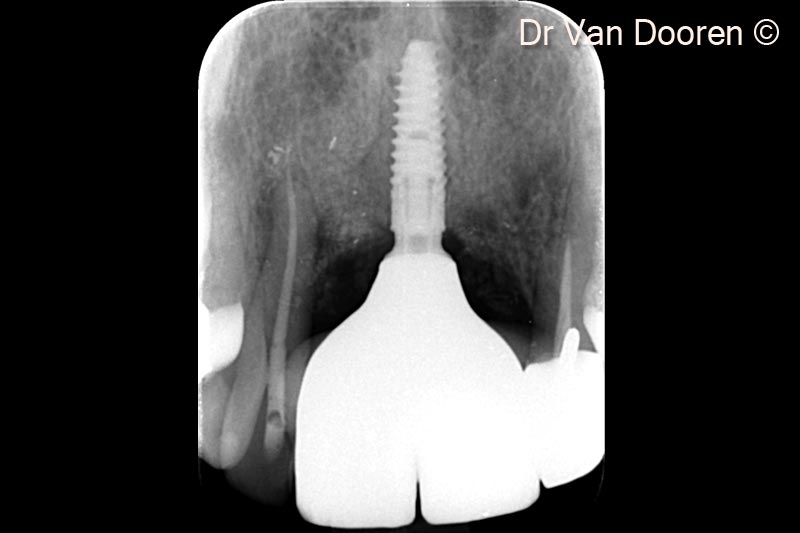
19. Final X-Ray. Note stable marginal bone levels around the implant and the nice integration of the graft (OsteoBiol® GTO®) in the peri-implant space
Attention please! The OsteoBiol® website contains information on Medical Devices, which may be dangerous for the patient health and safety if not used exclusively by medical professionals.