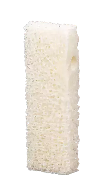
A female patient (70 years old) shows an atrophic posterior right mandible
- Sp-Block
- Evolution
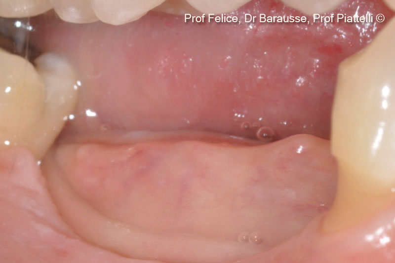
1. Pre-operative clinical image of an atrophic posterior right mandible
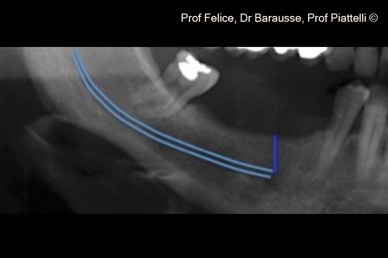
2. Pre-operative x-ray of the atrophic posterior right mandible
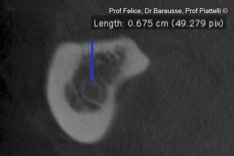
3. The CT cross section reveals the vertical crestal atrophy
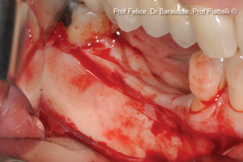
4. After performing a paracrestal incision in the buccal aspect, a flap is carefully elevated
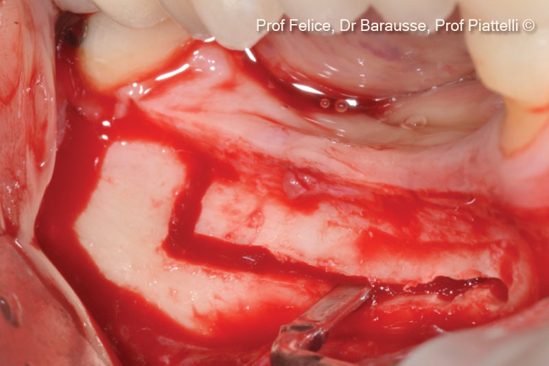
5. Horizontal and vertical osteotomy lines
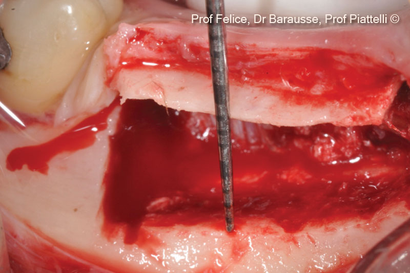
6. The osteotomized segment is raised coronally
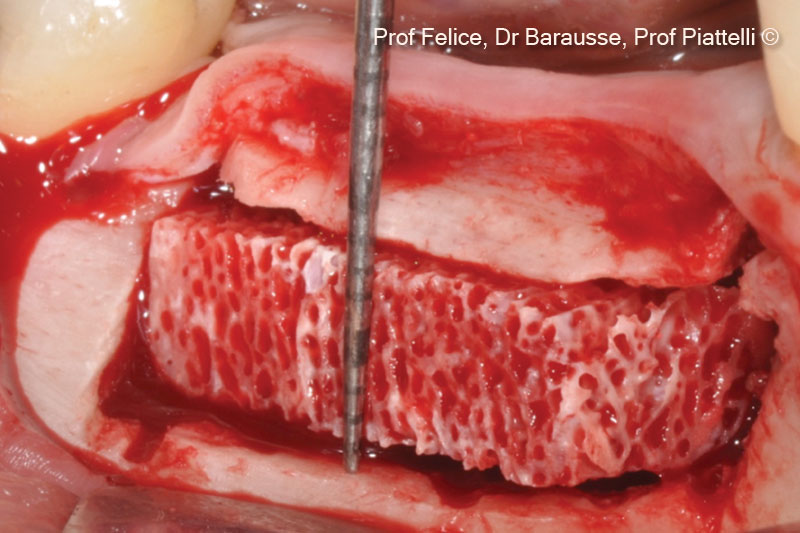
7. Sp-Block is placed in the obtained space
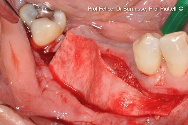
8. An Evolution membrane is applied on the vestibular surgical side
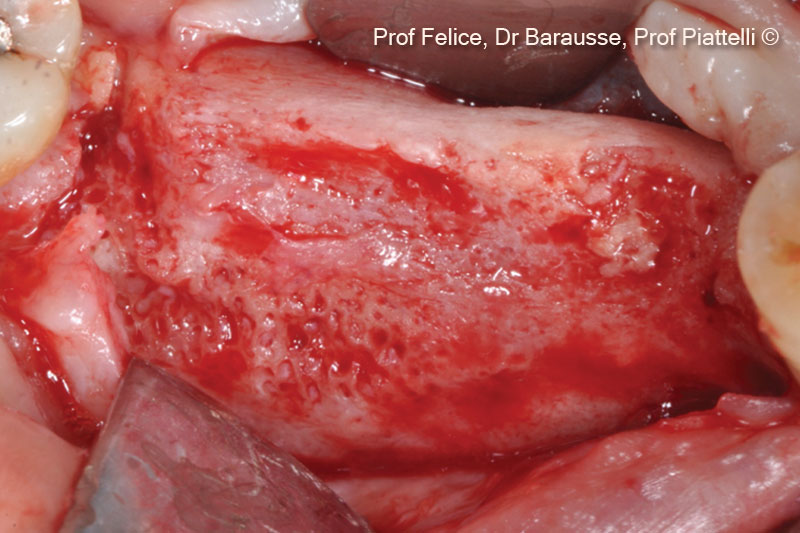
9. Clinical image of the re-entry surgery before implants positioning after 3 months
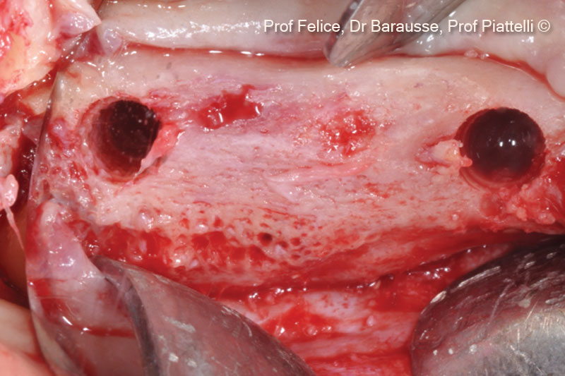
10. Implant tunnel preparations in the regenerated bone
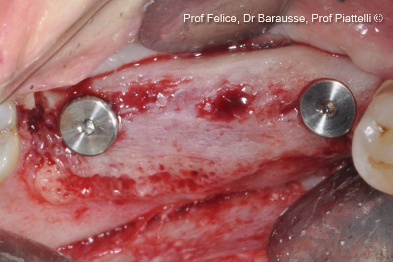
11. Two implants are placed during the re-entry surgery
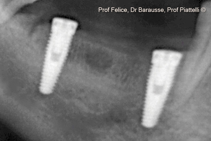
12. X-ray after implant positioning
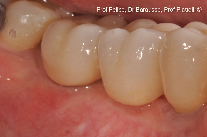
13. Clinical image of the definitive prosthesis after one year
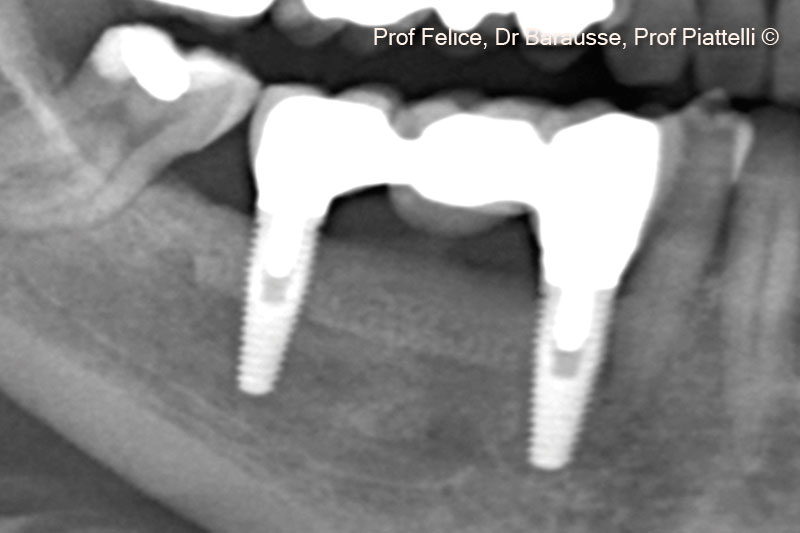
14. X-ray with definitive proshtesis after three years
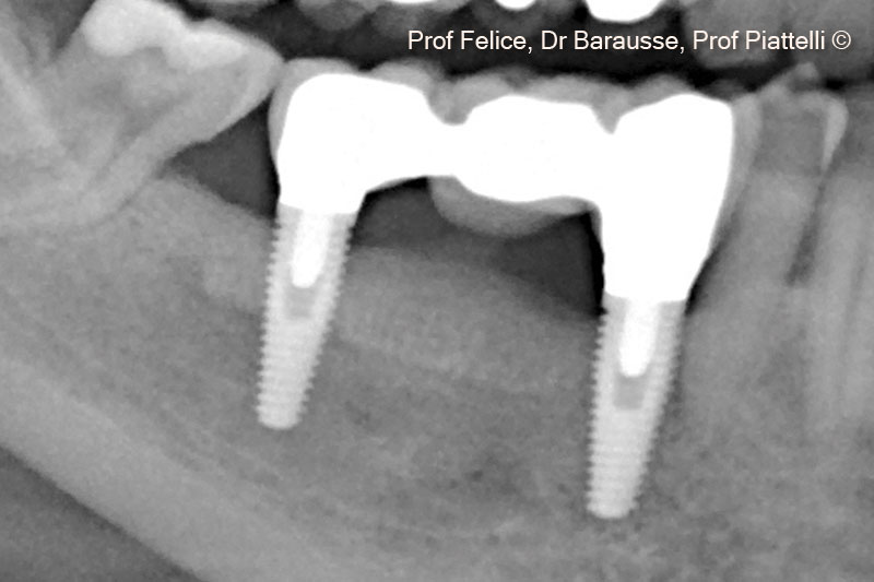
15. X-ray with definitive proshtesis after five years
